 Explore
 Validate
 Learn
Learn Western blot
 Immunocytochemistry
 Immunoprecipitation
Antibody data
- Antibody Data
- Antigen structure
- References [7]
- Comments [0]
- Validations
- Immunocytochemistry [1]
- Immunohistochemistry [1]
- Other assay [1]
Submit
Validation data
Reference
Comment
Report error
- Product number
- AHO1031 - Provider product page

- Provider
- Invitrogen Antibodies
- Product name
- PRAS40 Monoclonal Antibody (73P21)
- Antibody type
- Monoclonal
- Antigen
- Recombinant full-length protein
- Reactivity
- Human
- Host
- Mouse
- Isotype
- IgG
- Antibody clone number
- 73P21
- Vial size
- 100 μg
- Concentration
- 0.5 mg/mL
- Storage
- -20°C
Submitted references Location-specific inhibition of Akt reveals regulation of mTORC1 activity in the nucleus.
Combination of heat shock protein 90 and focal adhesion kinase inhibitors synergistically inhibits the growth of non-small cell lung cancer cells.
Characterization of the activity of the PI3K/mTOR inhibitor XL765 (SAR245409) in tumor models with diverse genetic alterations affecting the PI3K pathway.
Effects of adding exercise to a 16-week very low-calorie diet in obese, insulin-dependent type 2 diabetes mellitus patients.
Phosphorylation of PRAS40 on Thr246 by PKB/AKT facilitates efficient phosphorylation of Ser183 by mTORC1.
Insulin signalling to mTOR mediated by the Akt/PKB substrate PRAS40.
The proline-rich Akt substrate of 40 kDa (PRAS40) is a physiological substrate of mammalian target of rapamycin complex 1.
Zhou X, Zhong Y, Molinar-Inglis O, Kunkel MT, Chen M, Sun T, Zhang J, Shyy JY, Trejo J, Newton AC, Zhang J
Nature communications 2020 Nov 30;11(1):6088
Nature communications 2020 Nov 30;11(1):6088
Combination of heat shock protein 90 and focal adhesion kinase inhibitors synergistically inhibits the growth of non-small cell lung cancer cells.
Webber PJ, Park C, Qui M, Ramalingam SS, Khuri FR, Fu H, Du Y
Oncoscience 2015;2(9):765-776
Oncoscience 2015;2(9):765-776
Characterization of the activity of the PI3K/mTOR inhibitor XL765 (SAR245409) in tumor models with diverse genetic alterations affecting the PI3K pathway.
Yu P, Laird AD, Du X, Wu J, Won KA, Yamaguchi K, Hsu PP, Qian F, Jaeger CT, Zhang W, Buhr CA, Shen P, Abulafia W, Chen J, Young J, Plonowski A, Yakes FM, Chu F, Lee M, Bentzien F, Lam ST, Dale S, Matthews DJ, Lamb P, Foster P
Molecular cancer therapeutics 2014 May;13(5):1078-91
Molecular cancer therapeutics 2014 May;13(5):1078-91
Effects of adding exercise to a 16-week very low-calorie diet in obese, insulin-dependent type 2 diabetes mellitus patients.
Snel M, Gastaldelli A, Ouwens DM, Hesselink MK, Schaart G, Buzzigoli E, Frölich M, Romijn JA, Pijl H, Meinders AE, Jazet IM
The Journal of clinical endocrinology and metabolism 2012 Jul;97(7):2512-20
The Journal of clinical endocrinology and metabolism 2012 Jul;97(7):2512-20
Phosphorylation of PRAS40 on Thr246 by PKB/AKT facilitates efficient phosphorylation of Ser183 by mTORC1.
Nascimento EB, Snel M, Guigas B, van der Zon GC, Kriek J, Maassen JA, Jazet IM, Diamant M, Ouwens DM
Cellular signalling 2010 Jun;22(6):961-7
Cellular signalling 2010 Jun;22(6):961-7
Insulin signalling to mTOR mediated by the Akt/PKB substrate PRAS40.
Vander Haar E, Lee SI, Bandhakavi S, Griffin TJ, Kim DH
Nature cell biology 2007 Mar;9(3):316-23
Nature cell biology 2007 Mar;9(3):316-23
The proline-rich Akt substrate of 40 kDa (PRAS40) is a physiological substrate of mammalian target of rapamycin complex 1.
Oshiro N, Takahashi R, Yoshino K, Tanimura K, Nakashima A, Eguchi S, Miyamoto T, Hara K, Takehana K, Avruch J, Kikkawa U, Yonezawa K
The Journal of biological chemistry 2007 Jul 13;282(28):20329-39
The Journal of biological chemistry 2007 Jul 13;282(28):20329-39
No comments: Submit comment
Supportive validation
- Submitted by
- Invitrogen Antibodies (provider)
- Main image
- Experimental details
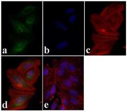
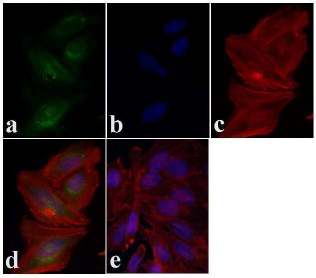
- Immunofluorescent analysis of PRAS40 was done on 70% confluent log phase HeLa cells. The cells were fixed with 4% paraformaldehyde for 15 minutes, permeabilized with 0.25% Triton X-100 for 10 minutes, and blocked with 5% BSA for 1 hour at room temperature. The cells were labeled with PRAS40 Mouse monoclonal Antibody (Product # AHO1031) at 2 µg/mL in 1% BSA and incubated for 3 hours at room temperature and then labeled with Alexa Fluor 488 Rabbit Anti-Mouse IgG Secondary Antibody (Product # A-11059) at a dilution of 1:400 for 30 minutes at room temperature (Panel a: green). Nuclei (Panel b: blue) were stained with SlowFade® Gold Antifade Mountant DAPI (Product # S36938). F-actin (Panel c: red) was stained with Alexa Fluor 594 Phalloidin (Product # A12381). Panel d is a merged image showing cytoplasmic localization. Panel e shows no primary antibody control. The images were captured at 20X magnification.
Supportive validation
- Submitted by
- Invitrogen Antibodies (provider)
- Main image

- Experimental details
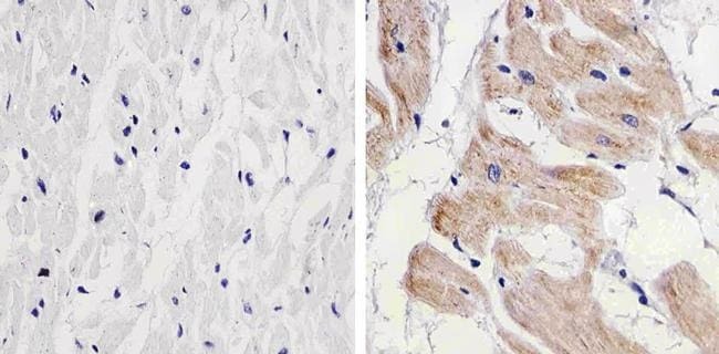
- Immunohistochemistry analysis of PRAS40 showing staining in the cytoplasm and nucleus of paraffin-embedded human heart tissue (right) compared to a negative control without primary antibody (left). To expose target proteins, antigen retrieval was performed using 10mM sodium citrate (pH 6.0), microwaved for 8-15 min. Following antigen retrieval, tissues were blocked in 3% H2O2-methanol for 15 min at room temperature, washed with ddH2O and PBS, and then probed with a PRAS40 monoclonal antibody (Product # AHO1031) diluted in 3% BSA-PBS at a dilution of 1:20 overnight at 4ºC in a humidified chamber. Tissues were washed extensively in PBST and detection was performed using an HRP-conjugated secondary antibody followed by colorimetric detection using a DAB kit. Tissues were counterstained with hematoxylin and dehydrated with ethanol and xylene to prep for mounting.
Supportive validation
- Submitted by
- Invitrogen Antibodies (provider)
- Main image
- Experimental details
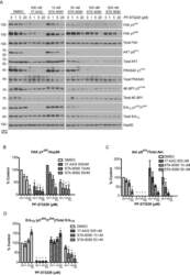
- Figure 4 Co-treatment of Hsp90 and FAK inhibitors reduces canonical survival signaling and induces apoptotic markers in H460 cells A) H460 cells treated for 24h with Hsp90 and FAK inhibitors were used for western blot analysis of FAK, Akt and Erk1/2 signaling. B-D) Quantifications of western blots from A. The phosphorylated proteins, FAK pY 397 (B), Akt pS 473 (C), Erk1/2 pT 202 /Y 204 (D), were normalized to Hsp90, total Akt and Erk1/2, respectively. Analyses were conducted by one-way ANOVA and Dunnett's multiple comparison post-test to compare all columns to control (* p
