Explore
 Validate
 Learn
Learn Western blot
Antibody data
- Antibody Data
- Antigen structure
- References [0]
- Comments [0]
- Validations
- Western blot [1]
- Immunohistochemistry [1]
- Flow cytometry [1]
Submit
Validation data
Reference
Comment
Report error
- Product number
- AF6836 - Provider product page

- Provider
- R&D Systems
- Product name
- Mouse Plexin B2 Antibody
- Antibody type
- Polyclonal
- Description
- Antigen Affinity-purified. Detects mouse Plexin B2 in direct ELISAs and Western blots. In direct ELISAs, approximately 9% cross-reactivity with recombinant human Plexin B2 is observed, and less than 1% cross-reactivity with recombinant mouse Plexin B3 is observed.
- Reactivity
- Mouse
- Host
- Sheep
- Conjugate
- Unconjugated
- Antigen sequence
B2RXS4- Isotype
- IgG
- Vial size
- 100 ug
- Concentration
- LYOPH
- Storage
- Use a manual defrost freezer and avoid repeated freeze-thaw cycles. 12 months from date of receipt, -20 to -70 °C as supplied. 1 month, 2 to 8 °C under sterile conditions after reconstitution. 6 months, -20 to -70 °C under sterile conditions after reconstitution.
No comments: Submit comment
Supportive validation
- Submitted by
- R&D Systems (provider)
- Main image
- Experimental details
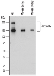
- Detection of Mouse Plexin B2 by Western Blot. Western blot shows lysates of M1 mouse myeloid leukemia cell line, mouse lung tissue, and mouse ovary tissue. PVDF membrane was probed with 0.5 µg/mL of Sheep Anti-Mouse Plexin B2 Antigen Affinity-purified Polyclonal Antibody (Catalog # AF6836) followed by HRP-conjugated Anti-Sheep IgG Secondary Antibody (Catalog # HAF016). Specific bands were detected for Plexin B2 at approximately 240 and 150 kDa (as indicated). This experiment was conducted under reducing conditions and using Immunoblot Buffer Group 1.

Supportive validation
- Submitted by
- R&D Systems (provider)
- Main image

- Experimental details
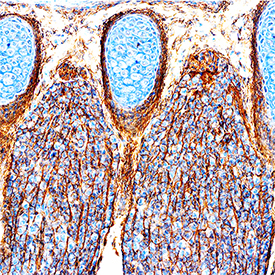
- Plexin B2 in Mouse Embryo. Plexin B2 was detected in immersion fixed frozen sections of mouse embryo (13 d.p.c.) using Sheep Anti-Mouse Plexin B2 Antigen Affinity-purified Polyclonal Antibody (Catalog # AF6836) at 0.6 µg/mL for 1 hour at room temperature followed by incubation with the Anti-Sheep IgG VisUCyte™ HRP Polymer Antibody (Catalog # VC006). Tissue was stained using DAB (brown) and counterstained with hematoxylin (blue). Specific staining was localized to developing central nervous system. View our protocol for IHC Staining with VisUCyte HRP Polymer Detection Reagents.
Supportive validation
- Submitted by
- R&D Systems (provider)
- Main image

- Experimental details
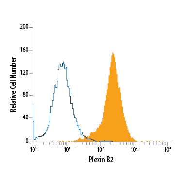
- Detection of Plexin B2 in RAW 264.7 Mouse Cell Line by Flow Cytometry. RAW 264.7 mouse monocyte/macrophage cell line was stained with Sheep Anti-Mouse Plexin B2 Antigen Affinity-purified Polyclonal Antibody (Catalog # AF6836, filled histogram) or isotype control antibody (Catalog # 5-001-A, open histogram), followed by Phycoerythrin-conjugated Anti-Sheep IgG Secondary Antibody (Catalog # F0126).