 Explore
 Validate
 Learn
Learn Western blot
Antibody data
- Antibody Data
- Antigen structure
- References [0]
- Comments [0]
- Validations
- Western blot [3]
- Immunohistochemistry [2]
Submit
Validation data
Reference
Comment
Report error
- Product number
- PA5-47721 - Provider product page

- Provider
- Invitrogen Antibodies
- Product name
- PLXNB2 Polyclonal Antibody
- Antibody type
- Polyclonal
- Antigen
- Recombinant full-length protein
- Description
- In direct ELISAs, less than 1% cross-reactivity with recombinant mouse (rm) Plexin A1, rmPlexin A2, recombinant human (rh) Plexin A4, rhPlexin B1, rhPlexin B3, rhPlexin C1 and rhPlexin D1 is observed. Reconstitute at 0.2 mg/mL in sterile PBS.
- Reactivity
- Human
- Host
- Sheep
- Isotype
- IgG
- Vial size
- 100 µg
- Concentration
- 0.2 mg/mL
- Storage
- -20° C, Avoid Freeze/Thaw Cycles
No comments: Submit comment
Supportive validation
- Submitted by
- Invitrogen Antibodies (provider)
- Main image

- Experimental details
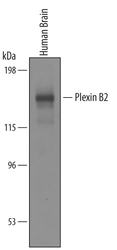
- Western blot analysis from lysates of human brain tissue. PVDF membrane was probed with 1 µg/mL of Sheep Anti-human Plexin B2 Antigen Affinity-purified Polyclonal Antibody (Product # PA5-47721) followed by HRP-conjugated Anti-Sheep IgG Secondary Antibody. A specific band was detected for Plexin B2 at approximately 170 kDa (as indicated). This experiment was conducted under reducing conditions.

- Submitted by
- Invitrogen Antibodies (provider)
- Main image
- Experimental details
- Western blot analysis of PLXNB2 in human brain tissue. Samples were incubated in PLXNB2 polyclonal antibody (Product # PA5-47721) using a dilution of 1 µg/mL followed by a HRP-conjugated Anti-Sheep IgG secondary antibody. A specific band was detected for Plexin B2 at approximately 170 kDa (as indicated). This experiment was conducted under reducing conditions.

- Submitted by
- Invitrogen Antibodies (provider)
- Main image

- Experimental details
- Knockout validation by Western blot analysis of PLXNB2 in lysates of HeLa human cervical epithelial carcinoma parental cell line and Plexin B2 knockout HeLa cell line (KO). Samples were incubated in PLXNB2 polyclonal antibody (Product # PA5-47721) using a dilution of 0.5 µg/mL followed by a HRP-conjugated Anti-Sheep IgG secondary antibody. A specific band was detected for Plexin B2 at approximately 170 kDa (as indicated) in the parental HeLa cell line, but is not detectable in knockout HeLa cell line. GAPDH is shown as a loading control. This experiment was conducted under reducing conditions.

Supportive validation
- Submitted by
- Invitrogen Antibodies (provider)
- Main image
- Experimental details
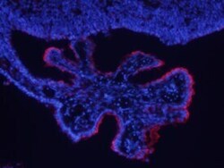
- Immunohistochemical analysis of PLXNB2 in perfusion fixed frozen sections of mouse brain (choroid plexus). Samples were incubated in PLXNB2 polyclonal antibody (Product # PA5-47721) using a dilution of 10 µg/mL overnight at 4 °C followed by NorthernLights™ 557-conjugated Anti-Sheep IgG Secondary Antibody (red) and counterstained with DAPI (blue).

- Submitted by
- Invitrogen Antibodies (provider)
- Main image
- Experimental details
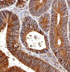
- Immunohistochemical analysis of PLXNB2 in immersion fixed paraffin-embedded sections of human colon cancer. Samples were incubated in PLXNB2 polyclonal antibody (Product # PA5-47721) using a dilution of 3 µg/mL overnight at 4 °C. Before incubation with the primary antibody tissue was subjected to heat-induced epitope retrieval using Antigen Retrieval Reagent-basic. Tissue was stained with the Anti-Sheep HRP-DAB Cell & Tissue Staining Kit (brown) and counterstained with hematoxylin (blue). Specific labeling was localized to the plasma membrane and cytoplasm of epithelial cells.
