Explore
 Validate
 Learn
Learn Western blot
Antibody data
- Antibody Data
- Antigen structure
- References [0]
- Comments [0]
- Validations
- Western blot [2]
- Immunocytochemistry [2]
- Immunohistochemistry [1]
Submit
Validation data
Reference
Comment
Report error
- Product number
- RQ4503 - Provider product page

- Provider
- NSJ Bioreagents
- Product name
- Phospho-Histone H2AX Antibody (pS139)
- Antibody type
- Monoclonal
- Description
- This highly specific Phospho-Histone H2AX antibody is suitable for use in Immunohistochemistry/Immunofluorescence/Immunocytochemistry/Western blot applications with human and mouse samples.
- Reactivity
- Human, Mouse
- Host
- Rabbit
- Conjugate
- Unconjugated
- Antibody clone number
- AbH41
- Vial size
- 100 ul
- Concentration
- Antibody in PBS with 0.02% sodium azide, 50% glycerol and 0.4-0.5mg/ml BSA
- Storage
- After reconstitution, the phospho-Histone H2AX antibody can be stored for up to one month at 4oC. For long-term, aliquot and store at -20oC. Avoid repeated freezing and thawing.
No comments: Submit comment
Supportive validation
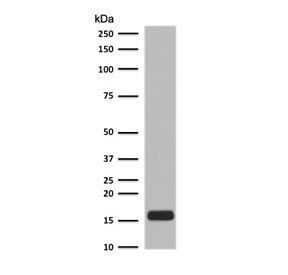
- Submitted by
- NSJ Bioreagents (provider)
- Main image

- Experimental details
- Western blot testing of etoposide-treated human Jurkat lysate with phospho-Histone H2AX antibody.

- Submitted by
- NSJ Bioreagents (provider)
- Main image

- Experimental details
- Western blot testing of etoposide-treated human Jurkat lysate with phospho-Histone H2AX antibody.
Supportive validation
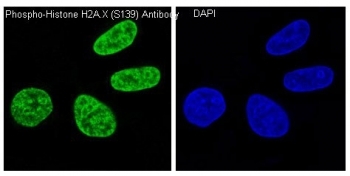
- Submitted by
- NSJ Bioreagents (provider)
- Main image

- Experimental details
- Immunofluorescent staining of human HeLa cells treated and untreated with H2O2 using phospho-Histone H2AX antibody (green) and DAPI nuclear stain (blue).

- Submitted by
- NSJ Bioreagents (provider)
- Main image

- Experimental details
- Immunofluorescent staining of human HeLa cells treated and untreated with H2O2 using phospho-Histone H2AX antibody (green) and DAPI nuclear stain (blue).
Supportive validation
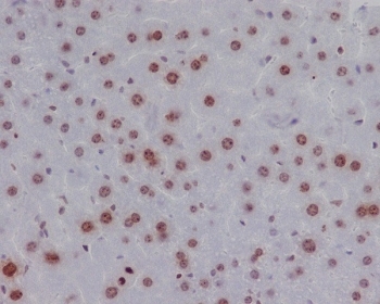
- Submitted by
- NSJ Bioreagents (provider)
- Main image
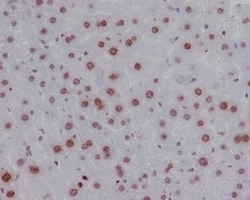
- Experimental details
- IHC testing of FFPE mouse liver with phospho-Histone H2AX antibody. HIER: boil tissue sections in pH6, 10mM citrate buffer, for 10-20 min followed by cooling at RT for 20 min.