Explore
 Validate
 Learn
Learn Western blot
Antibody data
- Antibody Data
- Antigen structure
- References [4]
- Comments [0]
- Validations
- Western blot [1]
- Immunocytochemistry [1]
Submit
Validation data
Reference
Comment
Report error
- Product number
- MAB3406 - Provider product page

- Provider
- R&D Systems
- Product name
- Human/Mouse/Rat Histone H2AX Antibody
- Antibody type
- Monoclonal
- Description
- Protein A or G purified from hybridoma culture supernatant. Detects human, mouse and rat Histone H2AX in Western blots.
- Reactivity
- Human, Mouse, Rat
- Host
- Mouse
- Conjugate
- Unconjugated
- Antigen sequence
P16104- Isotype
- IgG
- Antibody clone number
- 322105
- Vial size
- 50 ug
- Concentration
- LYOPH
- Storage
- Use a manual defrost freezer and avoid repeated freeze-thaw cycles. 12 months from date of receipt, -20 to -70 °C as supplied. 1 month, 2 to 8 °C under sterile conditions after reconstitution. 6 months, -20 to -70 °C under sterile conditions after reconstitution.
Submitted references Phenotypic Plasticity of Invasive Edge Glioma Stem-like Cells in Response to Ionizing Radiation.
Platelet glycoprotein VI and C-type lectin-like receptor 2 deficiency accelerates wound healing by impairing vascular integrity in mice.
miR-24-mediated knockdown of H2AX damages mitochondria and the insulin signaling pathway.
The formyl peptide receptor 1 exerts a tumor suppressor function in human gastric cancer by inhibiting angiogenesis.
Minata M, Audia A, Shi J, Lu S, Bernstock J, Pavlyukov MS, Das A, Kim SH, Shin YJ, Lee Y, Koo H, Snigdha K, Waghmare I, Guo X, Mohyeldin A, Gallego-Perez D, Wang J, Chen D, Cheng P, Mukheef F, Contreras M, Reyes JF, Vaillant B, Sulman EP, Cheng SY, Markert JM, Tannous BA, Lu X, Kango-Singh M, Lee LJ, Nam DH, Nakano I, Bhat KP
Cell reports 2019 Feb 12;26(7):1893-1905.e7
Cell reports 2019 Feb 12;26(7):1893-1905.e7
Platelet glycoprotein VI and C-type lectin-like receptor 2 deficiency accelerates wound healing by impairing vascular integrity in mice.
Wichaiyo S, Lax S, Montague SJ, Li Z, Grygielska B, Pike JA, Haining EJ, Brill A, Watson SP, Rayes J
Haematologica 2019 Aug;104(8):1648-1660
Haematologica 2019 Aug;104(8):1648-1660
miR-24-mediated knockdown of H2AX damages mitochondria and the insulin signaling pathway.
Jeong JH, Cheol Kang Y, Piao Y, Kang S, Pak YK
Experimental & molecular medicine 2017 Apr 7;49(4):e313
Experimental & molecular medicine 2017 Apr 7;49(4):e313
The formyl peptide receptor 1 exerts a tumor suppressor function in human gastric cancer by inhibiting angiogenesis.
Prevete N, Liotti F, Visciano C, Marone G, Melillo RM, de Paulis A
Oncogene 2015 Jul;34(29):3826-38
Oncogene 2015 Jul;34(29):3826-38
No comments: Submit comment
Supportive validation
- Submitted by
- R&D Systems (provider)
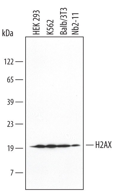
- Main image
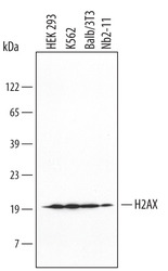
- Experimental details
- Detection of Human/Mouse/Rat Histone H2AX by Western Blot. Western blot shows lysates of HEK293 human embryonic kidney cell line, K562 human chronic myelogenous leukemia cell line, Balb/3T3 mouse embryonic fibroblast cell line, and Nb2-11 rat lymphoma cell line. PVDF membrane was probed with 0.5 µg/mL of Mouse Anti-Human/Mouse/Rat Histone H2AX Monoclonal Antibody (Catalog # MAB3406) followed by HRP-conjugated Anti-Mouse IgG Secondary Antibody (Catalog # HAF007). A specific band was detected for Histone H2AX at approximately 20 kDa (as indicated). This experiment was conducted under reducing conditions and using Immunoblot Buffer Group 1.
Supportive validation
- Submitted by
- R&D Systems (provider)
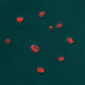
- Main image
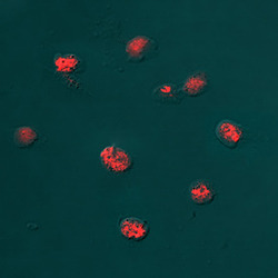
- Experimental details
- Histone H2AX in HeLa Human Cell Line. Histone H2AX was detected in immersion fixed HeLa human cervical epithelial carcinoma cell line using Mouse Anti-Human/Mouse/Rat Histone H2AX Monoclonal Antibody (Catalog # MAB3406) at 8 µg/mL for 3 hours at room temperature. Cells were stained using the Northern-Lights™ 557-conjugated Anti-Mouse IgG Secondary Antibody (red; Catalog # NL007) and counterstained with DAPI (blue). Specific staining was localized to nuclei. View our protocol for Fluorescent ICC Staining of Cells on Coverslips.