Explore
 Validate
 Learn
Learn Western blot
Antibody data
- Antibody Data
- Antigen structure
- References [0]
- Comments [0]
- Validations
- Western blot [3]
- Immunohistochemistry [3]
Submit
Validation data
Reference
Comment
Report error
- Product number
- PA5-30416 - Provider product page

- Provider
- Invitrogen Antibodies
- Product name
- HP1 beta Polyclonal Antibody
- Antibody type
- Polyclonal
- Antigen
- Recombinant protein fragment
- Description
- Recommended positive controls: HeLa, HepG2, HCT116.
- Concentration
- 0.54 mg/mL
No comments: Submit comment
Supportive validation
- Submitted by
- Invitrogen Antibodies (provider)
- Main image
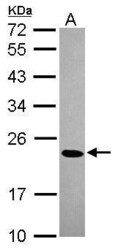
- Experimental details
- Western Blot using HP1 beta Polyclonal Antibody (Product # PA5-30416). Sample (30 µg of whole cell lysate). Lane A: HeLa. 12% SDS PAGE. HP1 beta Polyclonal Antibody (Product # PA5-30416) diluted at 1:1,000.

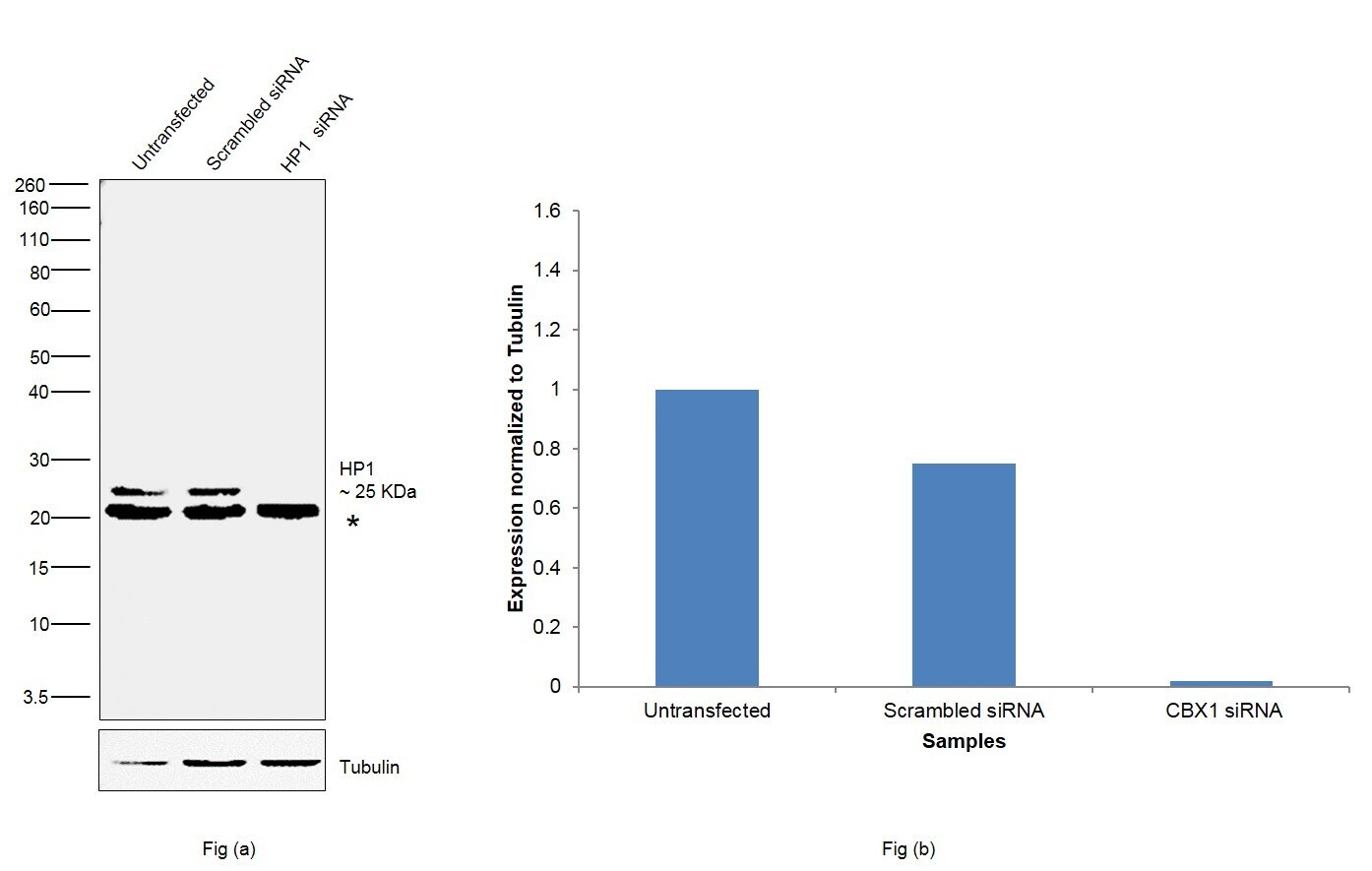
- Submitted by
- Invitrogen Antibodies (provider)
- Main image

- Experimental details
- Knockdown of HP1 was achieved by transfecting MCF7 with HP1 specific siRNAs (Silencer® select Product # s21549). Western blot analysis (Fig. a) was performed using whole cell extracts from the HP1 knockdown cells (lane 3), non-specific scrambled siRNA transfected cells (lane 2) and untransfected cells (lane 1). The blot was probed with HP1 Monoclonal Antibody (Product # PA5-30416, 1:1000 dilution) and Goat anti-Rabbit IgG (H+L) Superclonal™ Secondary Antibody, HRP (Product # A27036, 1:4000 dilution) . Densitometric analysis of this western blot is shown in histogram (Fig. b). Decrease in signal upon siRNA mediated knock down confirms that antibody is specific to HP1.
- Submitted by
- Invitrogen Antibodies (provider)
- Main image
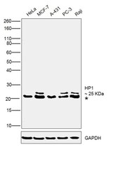
- Experimental details
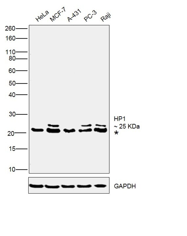
- Western blot was performed using Anti- HP1 Polyclonal Antibody(Product # PA5-30416) and a 25 kDa band corresponding to HP1 was observed along with uncharacterized band (*) across the cell lines and tissues tested. Modified whole cell extracts (1% SDS) (30ug lysate) of HeLa (Lane 1), MCF-7 (Lane 2), A-431 (Lane 3), PC-3 (Lane 4) and Raji (Lane 5) were electrophoresed using Novex® NuPAGE® 4-12 % Bis-Tris gel (Product # NP0322BOX). Resolved proteins were then transferred onto a nitrocellulose membrane (Product # IB23001) by iBlot® 2 Dry Blotting System (Product # IB21001). The blot was probed with the primary antibody (1:1000 dilution) and detected by chemiluminescence with Goat anti-Rabbit IgG (H+L) Superclonal™ Recombinant Secondary Antibody, HRP (Product # A27036, 1:4000 dilution) using the iBright FL 1000 (Product # A32752). Chemiluminescent detection was performed using Novex® ECL Chemiluminescent Substrate Reagent Kit (Product # WP20005).
Supportive validation
- Submitted by
- Invitrogen Antibodies (provider)
- Main image
- Experimental details
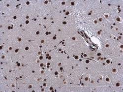
- CBX1/HP1 beta antibody detects CBX1/HP1 beta protein at nucleus in mouse brain by immunohistochemical analysis. Sample: Paraffin-embedded mouse brain. CBX1/HP1 beta antibody (Product # PA5-30416) diluted at 1:500. Antigen Retrieval: Citrate buffer, pH 6.0, 15 min.
- Submitted by
- Invitrogen Antibodies (provider)
- Main image
- Experimental details
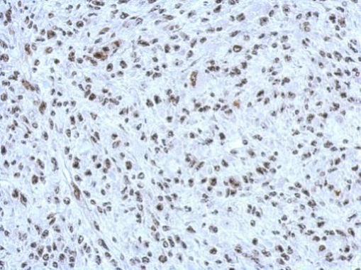
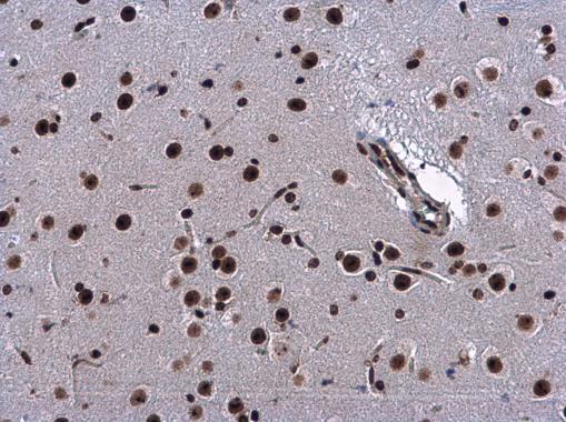
- Immunohistochemical analysis of paraffin-embedded 59T xenograft, using CBX1/HP1 beta (Product # PA5-30416) antibody at 1:500 dilution. Antigen Retrieval: EDTA based buffer, pH 8.0, 15 min.

- Submitted by
- Invitrogen Antibodies (provider)
- Main image

- Experimental details
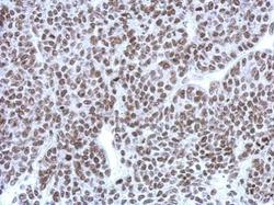
- Immunohistochemical analysis of paraffin-embedded C2C12 xenograft, using CBX1/HP1 beta (Product # PA5-30416) antibody at 1:500 dilution. Antigen Retrieval: EDTA based buffer, pH 8.0, 15 min.