Explore
 Validate
 Learn
LearnPA3-16744
antibody from Invitrogen Antibodies
Targeting: CADM1
BL2, IGSF4, IGSF4A, Necl-2, NECL2, RA175, ST17, SYNCAM, SYNCAM1, TSLC1
 Western blot
 Other assay
Antibody data
- Antibody Data
- Antigen structure
- References [2]
- Comments [0]
- Validations
- Other assay [4]
Submit
Validation data
Reference
Comment
Report error
- Product number
- PA3-16744 - Provider product page

- Provider
- Invitrogen Antibodies
- Product name
- CADM1 Polyclonal Antibody
- Antibody type
- Polyclonal
- Antigen
- Synthetic peptide
- Description
- Suggested positive control: rat brain.
- Reactivity
- Human, Rat
- Host
- Rabbit
- Isotype
- IgG
- Vial size
- 100 μL
- Concentration
- Conc. Not Determined
- Storage
- Store at 4°C short term. For long term storage, store at -20°C, avoiding freeze/thaw cycles.
Submitted references Redundant Postsynaptic Functions of SynCAMs 1-3 during Synapse Formation.
MPP2 is a postsynaptic MAGUK scaffold protein that links SynCAM1 cell adhesion molecules to core components of the postsynaptic density.
Fowler DK, Peters JH, Williams C, Washbourne P
Frontiers in molecular neuroscience 2017;10:24
Frontiers in molecular neuroscience 2017;10:24
MPP2 is a postsynaptic MAGUK scaffold protein that links SynCAM1 cell adhesion molecules to core components of the postsynaptic density.
Rademacher N, Schmerl B, Lardong JA, Wahl MC, Shoichet SA
Scientific reports 2016 Oct 19;6:35283
Scientific reports 2016 Oct 19;6:35283
No comments: Submit comment
Supportive validation
- Submitted by
- Invitrogen Antibodies (provider)
- Main image
- Experimental details
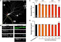
- Figure 2 SynCAM1-3 function redundantly to set synapse density and size. (A) Representative 60x confocal microscopy image of cultured rat hippocampal neurons at 14 DIV treated with sub-saturating lentivirus carrying Scrambled1-3 amiRNAs linked to nlsGFP, and very low titer memGFP lentivirus. An individual pyramidal cell co-labeled by memGFP and nlsGFP is marked by a red arrowhead. White arrowheads mark nuclei in the field of view only expressing nlsGFP. Scale, 50 mum. (B) Sample images of automated puncta and synapse detection from co-immunostaining for presynaptic protein Synapsin 1 and postsynaptic protein PSD-95 following manual selection of an individual dendrite segment from the image in (A) . Synapses are defined as the co-localized regions of pre- and postsynaptic puncta (white areas in merged image). Scale, 10 mum. (C) Average synapse density and (D) average synapse puncta area from 13 to 15 DIV neurons co-transduced with memGFP and sub-saturating amounts of the SynCAM amiRNAs listed or control Scrambled1-3 amiRNAs. n = cells/isolations is listed on the bars. SKD, single knockdown; DKD, double knockdown; TKD, triple knockdown. * p < 0.05, ** p < 0.01, one-way analyses of variance (ANOVA) with Tukey's post hoc pairwise comparisons on cell average values. Error bars, SEM.

- Submitted by
- Invitrogen Antibodies (provider)
- Main image
- Experimental details
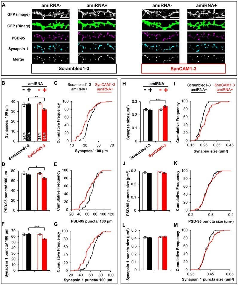
- Figure 4 MEDLR shows synaptic phenotypes are due to postsynaptic SynCAM1-3 depletion. (A) Representative images of memGFP-labled basal dendrites from amiRNA- and amiRNA+ neurons at 15 DIV in cultures transduced with sub-saturating amounts of Scrambled1-3 or SynCAM1-3 amiRNAs. Automated puncta detection for PSD-95 and Synapsin 1 immunostaining and synapses (merge) was performed after manual selection of GFP masks. Scale, 5 mum. (B,D,F) Average dendritic puncta density as indicated on graph of amiRNA- and amiRNA+ neurons and (C,E,G) corresponding cumulative puncta density distribution plot (%) of amiRNA+ neurons from cultures transduced with Scrambled1-3 or SynCAM1-3 amiRNAs. (H,J,L) Average puncta area as indicated on graph of amiRNA- and amiRNA+ neurons and (I,K,M) corresponding cumulative puncta area distribution plot (%) of amiRNA+ neurons from cultures transduced with Scrambled1-3 or SynCAM1-3 amiRNAs. n = number of cells/isolations as indicated on bars from 13 to 15 DIV cultures. * p < 0.05, ** p < 0.01, *** p < 0.001, t test on cell average values. Cumulative distribution plots represent cell average values. Error bars, SEM.
- Submitted by
- Invitrogen Antibodies (provider)
- Main image
- Experimental details
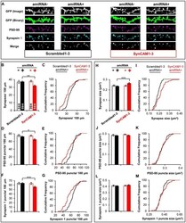
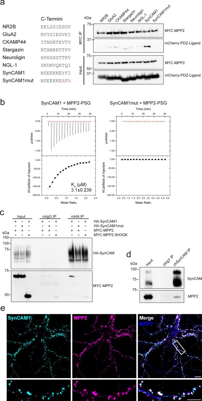
- Figure 3 MPP2 binds directly to the synaptic adhesion molecule SynCAM1. ( a ) Targeted identification of putative MPP2 PDZ domain interactors: The 10 C-terminal amino acids of known synaptic PDZ-ligand proteins (see table on the left) were fused to the monomeric mCherry tag and tested for interaction with MYC-tagged MPP2 in coimmunoprecipitation experiments. Pulldown control (MYC-MPP2) and coprecipitated proteins (mCherry-PDZ-Ligand) were detected by western blot (upper panels); input controls are shown below. For uncropped blots, see Supplementary Fig. S3 . ( b ) Isothermal titration calorimetry measurements with 350 muM mCherry-SynCAM1 and SynCAM1mut, respectively, injected to 55 muM MPP2-PSG module revealed a robust binding of the SynCAM1 C-terminus with a K d of 3.1 +- 0.239 muM (left panels). Results for the mutated SynCAM1 C-terminus control are shown on the right. ( c ) Full-length HA-tagged SynCAM1 (HA-SynCAM1) and MYC-tagged MPP2 (MYC-MPP2), HA-tagged mutant SynCAM1 (HA-SynCAM1mut) and MYC-MPP2, or HA-SynCAM1 and MYC-tagged MPP2 SH3-GK (MYC-MPP2-SH3GK) were coexpressed in COS7 cells and immunoprecipitated with alphaHA (mHA IP) or mouse IgGs (mIgG IP) as a negative control. Pulldown controls are shown (HA-SynCAM); coprecipitated proteins are detected by western blot with alphaMYC antibody below. For uncropped blots, see Supplementary Fig. S3 . ( d ) Immunoprecipitation of SynCAM1 (using a chicken SynCAM1-Biotin antibody or chicken IgYs as a negative control) from a cr
- Submitted by
- Invitrogen Antibodies (provider)
- Main image

- Experimental details
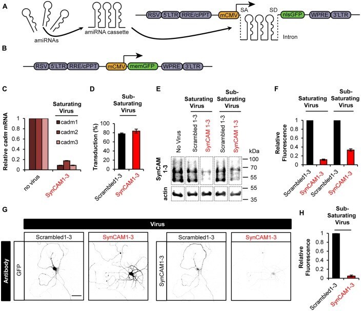
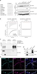
- Figure 1 Sub-saturating lentiviral artificial microRNA (amiRNA) transduction potently knocks down synaptic cell adhesion molecules 1-3 (SynCAM1-3) in amiRNA+ cells while keeping a subpopulation of wild type (WT) amiRNA- cells. (A) Lentiviral knockdown (KD) vectors were generated with a minimal CMV (mCMV) promoter, intronic amiRNAs, and nlsGFP. RSV, Rous sarcoma virus promoter; LTR, long-terminal repeat; RRE, Rev-response element; cPPT, central polypurine tract; WPRE, woodchuck hepatitis virus posttranscriptional regulatory element; SA, splice acceptor; SD, splice donor. (B) memGFP-expressing lentiviral vector for dendrite labeling. (C) Comparison of cadm1-3 mRNA levels at 14-15 days in vitro (DIV) by quantitative reverse transcription polymerase chain reaction (qRT-PCR) in cultured rat hippocampal neurons using saturating viral titers relative to levels in uninfected control cultures. n = 2 experiments. (D) Transduction rate of cultured neurons at 14-15 DIV using sub-saturating viral titers. n = 2 experiments. (E) Representative blots and (F) relative SynCAM1-3 protein levels at 13-15 DIV measured by quantitative western blotting of cultured neurons following indicated amiRNA lentiviral treatments at saturating or sub-saturating viral titers compared to Scrambled1-3 amiRNA control treatments. Actin was used as a loading control. n = 4 (saturating) or n = 2 (sub-saturating) experiments. A single gel image of the same intensity was cropped as marked by dotted lines and rearrang