Explore
 Validate
 Learn
Learn Western blot
Antibody data
- Antibody Data
- Antigen structure
- References [3]
- Comments [0]
- Validations
- Western blot [6]
- Immunocytochemistry [2]
- Immunohistochemistry [9]
Submit
Validation data
Reference
Comment
Report error
- Product number
- GTX115549 - Provider product page

- Provider
- GeneTex
- Proper citation
- GeneTex Cat#GTX115549, RRID:AB_10624805
- Product name
- Histone H3.3B antibody
- Antibody type
- Polyclonal
- Reactivity
- Human, Mouse, Rat
- Host
- Rabbit
Submitted references Placental miR-340 mediates vulnerability to activity based anorexia in mice.
Role of high mobility group box 1 (HMGB1) in SCA17 pathogenesis.
Aqueous Extract of Paeonia lactiflora and Paeoniflorin as Aggregation Reducers Targeting Chaperones in Cell Models of Spinocerebellar Ataxia 3.
Schroeder M, Jakovcevski M, Polacheck T, Drori Y, Luoni A, Röh S, Zaugg J, Ben-Dor S, Albrecht C, Chen A
Nature communications 2018 Apr 23;9(1):1596
Nature communications 2018 Apr 23;9(1):1596
Role of high mobility group box 1 (HMGB1) in SCA17 pathogenesis.
Lee LC, Chen CM, Wang PR, Su MT, Lee-Chen GJ, Chang CY
PloS one 2014;9(12):e115809
PloS one 2014;9(12):e115809
Aqueous Extract of Paeonia lactiflora and Paeoniflorin as Aggregation Reducers Targeting Chaperones in Cell Models of Spinocerebellar Ataxia 3.
Chang KH, Chen WL, Lee LC, Lin CH, Kung PJ, Lin TH, Wu YC, Wu YR, Chen YC, Lee-Chen GJ, Chen CM
Evidence-based complementary and alternative medicine : eCAM 2013;2013:471659
Evidence-based complementary and alternative medicine : eCAM 2013;2013:471659
No comments: Submit comment
Supportive validation
- Submitted by
- GeneTex (provider)
- Main image
- Experimental details
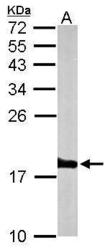
- Sample (50 ?g of whole cell lysate) A: mouse brain 15% SDS PAGE GTX115549 diluted at 1:1000 The HRP-conjugated anti-rabbit IgG antibody (GTX213110-01) was used to detect the primary antibody.

- Submitted by
- GeneTex (provider)
- Main image
- Experimental details
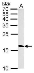
- Histone H3.3B antibody detects H3F3B protein by western blot analysis.A. 50 ?g rat brain lysate/extract15% SDS-PAGEHistone H3.3B antibody (GTX115549) dilution: 1:1000 The HRP-conjugated anti-rabbit IgG antibody (GTX213110-01) was used to detect the primary antibody.

- Submitted by
- GeneTex (provider)
- Main image

- Experimental details
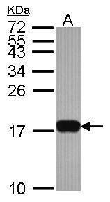
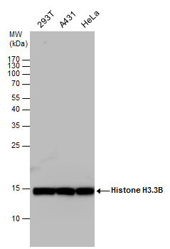
- Sample (30 ug of whole cell lysate) A: Hela 15% SDS PAGE GTX115549 diluted at 1:1000
- Validation comment
- WB
- Submitted by
- GeneTex (provider)
- Main image
- Experimental details
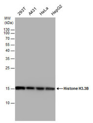
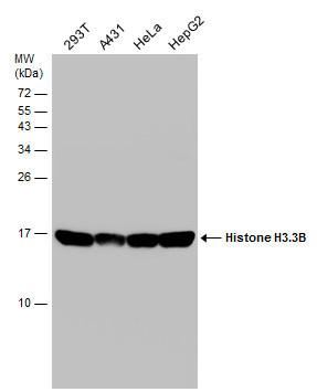
- Histone H3.3B antibody detects Histone H3.3B protein by western blot analysis. Various whole cell extracts (30 ?g) were separated by 15% SDS-PAGE, and the membrane was blotted with Histone H3.3B antibody (GTX115549) diluted by 1:10000.
- Validation comment
- WB
- Submitted by
- GeneTex (provider)
- Main image
- Experimental details
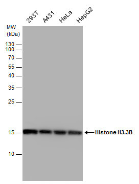
- Histone H3.3B antibody detects Histone H3.3B protein by western blot analysis. Various whole cell extracts (30 ?g) were separated by 15% SDS-PAGE, and the membrane was blotted with Histone H3.3B antibody (GTX115549) diluted at a dilution of 1:10000. The HRP-conjugated anti-rabbit IgG antibody (GTX213110-01) was used to detect the primary antibody.
- Submitted by
- GeneTex (provider)
- Main image

- Experimental details
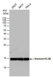
- Various whole cell extracts (30 ?g) were separated by 15% SDS-PAGE, and the membrane was blotted with Histone H3.3B antibody (GTX115549) diluted at 1:10000.
Supportive validation
- Submitted by
- GeneTex (provider)
- Main image

- Experimental details
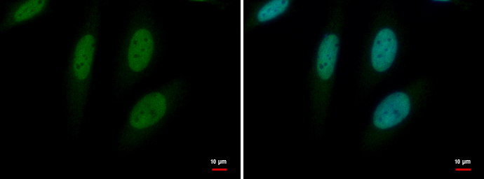
- Histone H3.3B antibody detects Histone H3.3B protein at nucleus by immunofluorescent analysis.Sample: HeLa cells were fixed in 4% paraformaldehyde at RT for 15 min.Green: Histone H3.3B protein stained by Histone H3.3B antibody (GTX115549) diluted at 1:500.Blue: Hoechst 33342 staining.
- Submitted by
- GeneTex (provider)
- Main image
- Experimental details
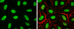
- Histone H3.3B antibody detects Histone H3.3B protein at nucleus by immunofluorescent analysis.Sample: HeLa cells were fixed in 4% paraformaldehyde at RT for 15 min.Green: Histone H3.3B protein stained by Histone H3.3B antibody (GTX115549) diluted at 1:500.Red: Phalloidin, a cytoskeleton marker, diluted at 1:100.

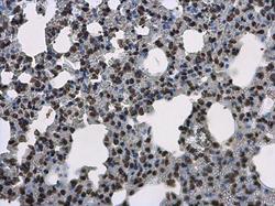
Supportive validation
- Submitted by
- GeneTex (provider)
- Main image
- Experimental details
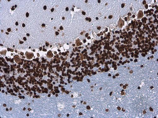
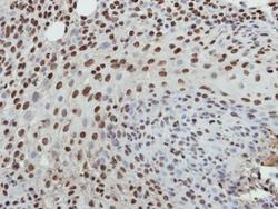
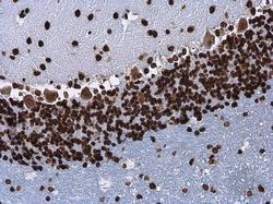
- Immunohistochemical analysis of paraffin-embedded HSC3 xenograft, using Histone H3.3B(GTX115549) antibody at 1:500 dilution.

- Submitted by
- GeneTex (provider)
- Main image
- Experimental details
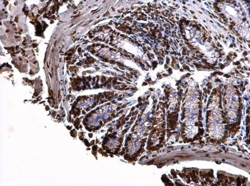
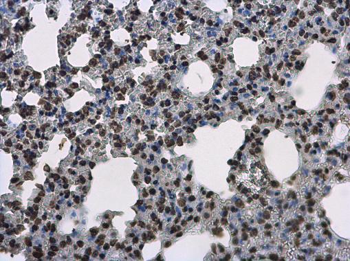
- Histone H3.3B antibody detects Histone H3.3B protein at nucleus on mouse colon by immunohistochemical analysis. Sample: Paraffin-embedded mouse colon. Histone H3.3B antibody (GTX115549) dilution: 1:500.
- Submitted by
- GeneTex (provider)
- Main image

- Experimental details
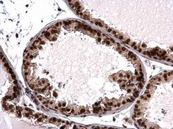
- Histone H3.3B antibody detects Histone H3.3B protein at nucleus on mouse prostate by immunohistochemical analysis. Sample: Paraffin-embedded mouse prostate. Histone H3.3B antibody (GTX115549) dilution: 1:500.
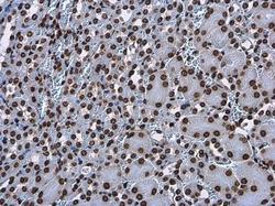
- Submitted by
- GeneTex (provider)
- Main image
- Experimental details
- Histone H3.3B antibody detects Histone H3.3B protein at nucleus on mouse lung by immunohistochemical analysis. Sample: Paraffin-embedded mouse lung. Histone H3.3B antibody (GTX115549) diluted at 1:500.
- Submitted by
- GeneTex (provider)
- Main image
- Experimental details
- Histone H3.3B antibody detects Histone H3.3B protein at nucleus on mouse spinal cord by immunohistochemical analysis. Sample: Paraffin-embedded mouse spinal cord. Histone H3.3B antibody (GTX115549) diluted at 1:500.
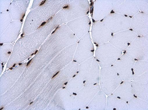
- Submitted by
- GeneTex (provider)
- Main image
- Experimental details
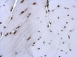
- Histone H3.3B antibody detects Histone H3.3B protein at nucleus in mouse muscle by immunohistochemical analysis. Sample: Paraffin-embedded mouse muscle. Histone H3.3B antibody (GTX115549) diluted at 1:500.
- Submitted by
- GeneTex (provider)
- Main image
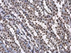
- Experimental details
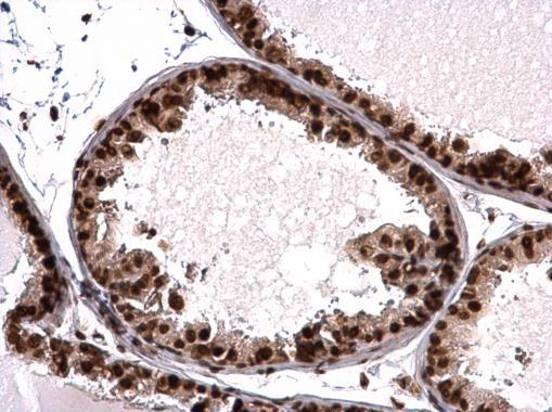
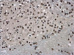
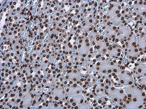
- Histone H3.3B antibody detects Histone H3.3B protein at nucleus in rat kidney by immunohistochemical analysis. Sample: Paraffin-embedded rat kidney. Histone H3.3B antibody (GTX115549) diluted at 1:500.
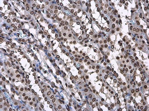
- Submitted by
- GeneTex (provider)
- Main image
- Experimental details
- Histone H3.3B antibody detects Histone H3.3B protein at nucleus in mouse kidney by immunohistochemical analysis. Sample: Paraffin-embedded mouse kidney. Histone H3.3B antibody (GTX115549) diluted at 1:500.
- Submitted by
- GeneTex (provider)
- Main image
- Experimental details
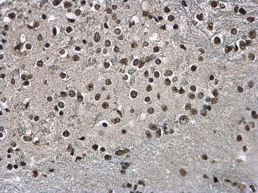
- Histone H3.3B antibody detects Histone H3.3B protein at nucleus in rat brain by immunohistochemical analysis. Sample: Paraffin-embedded rat brain. Histone H3.3B antibody (GTX115549) diluted at 1:500.