 Explore
 Validate
 Learn
Learn Western blot
 ELISA
ELISA Immunocytochemistry
Antibody data
- Antibody Data
- Antigen structure
- References [0]
- Comments [0]
- Validations
- Western blot [1]
- Immunocytochemistry [3]
Submit
Validation data
Reference
Comment
Report error
- Product number
- LS-C675332 - Provider product page

- Provider
- LSBio
- Product name
- H3F3B Antibody LS-C675332
- Antibody type
- Monoclonal
- Description
- Affinity chromatography
- Reactivity
- Human
- Host
- Rabbit
- Isotype
- IgG
- Storage
- Upon receipt, store at -20°C or -80°C. Avoid repeated freeze.
No comments: Submit comment
Enhanced validation
- Submitted by
- LSBio (provider)
- Enhanced method
- Genetic validation
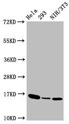
- Main image

- Experimental details
- Western Blot Positive WB detected in:Hela whole cell lysate, 293 whole cell lysate, NIH/3T3 whole cell lysate All Lanes:Phospho-Histone H3 (T3) antibody at 1.41µg/ml Secondary Goat polyclonal to rabbit IgG at 1/50000 dilution Predicted band size: 16 KDa Observed band size: 16 KDa
Supportive validation
- Submitted by
- LSBio (provider)
- Enhanced method
- Genetic validation
- Main image
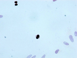
- Experimental details
- Immunocytochemistry analysis diluted at 1:100 and staining in Hela cells performed on a Leica BondTM system. After dewaxing and hydration, antigen retrieval was mediated by high pressure in a citrate buffer (pH 6.0). Section was blocked with 10% normal Goat serum 30min at RT. Then primary antibody (1% BSA) was incubated at 4°C overnight. The primary is detected by a biotinylated Secondary antibody and visualized using an HRP conjugated SP system.

- Submitted by
- LSBio (provider)
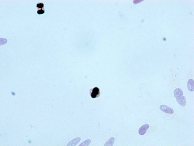
- Main image

- Experimental details
- Immunocytochemistry analysis diluted at 1:100 and staining in Hela cells performed on a Leica BondTM system. After dewaxing and hydration, antigen retrieval was mediated by high pressure in a citrate buffer (pH 6.0). Section was blocked with 10% normal Goat serum 30min at RT. Then primary antibody (1% BSA) was incubated at 4°C overnight. The primary is detected by a biotinylated Secondary antibody and visualized using an HRP conjugated SP system.
- Submitted by
- LSBio (provider)
- Main image

- Experimental details
- Immunocytochemistry analysis diluted at 1:100 and staining in Hela cells performed on a Leica BondTM system. After dewaxing and hydration, antigen retrieval was mediated by high pressure in a citrate buffer (pH 6.0). Section was blocked with 10% normal Goat serum 30min at RT. Then primary antibody (1% BSA) was incubated at 4°C overnight. The primary is detected by a biotinylated Secondary antibody and visualized using an HRP conjugated SP system.
