Explore
 Validate
 Learn
Learn Western blot
 ELISA
ELISA Immunocytochemistry
 Radioimmunoassay
Antibody data
- Antibody Data
- Antigen structure
- References [0]
- Comments [0]
- Validations
- Western blot [1]
- ELISA [9]
- Immunohistochemistry [2]
Submit
Validation data
Reference
Comment
Report error
- Product number
- LS-C355402 - Provider product page

- Provider
- LSBio
- Product name
- APOA1 / Apolipoprotein A 1 Antibody (clone 513) LS-C355402
- Antibody type
- Monoclonal
- Description
- Ion exchange chromatography
- Reactivity
- Human
- Host
- Mouse
- Isotype
- IgG
- Antibody clone number
- 513
- Storage
- Short term: store at 4°C. Long term: aliquot and store at -20°C. Avoid freeze-thaw cycles.
No comments: Submit comment
Enhanced validation
- Submitted by
- LSBio (provider)
- Enhanced method
- Genetic validation
- Main image
- Experimental details
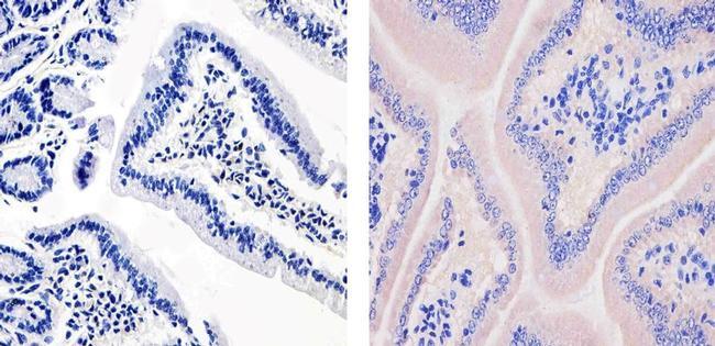
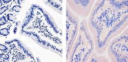
- Apolipoprotein A-1 Immunohistochemistry
Supportive validation
- Submitted by
- LSBio (provider)
- Enhanced method
- Genetic validation
- Main image
- Experimental details
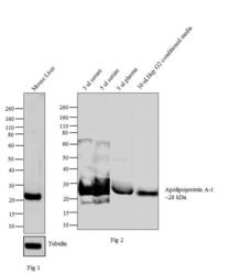
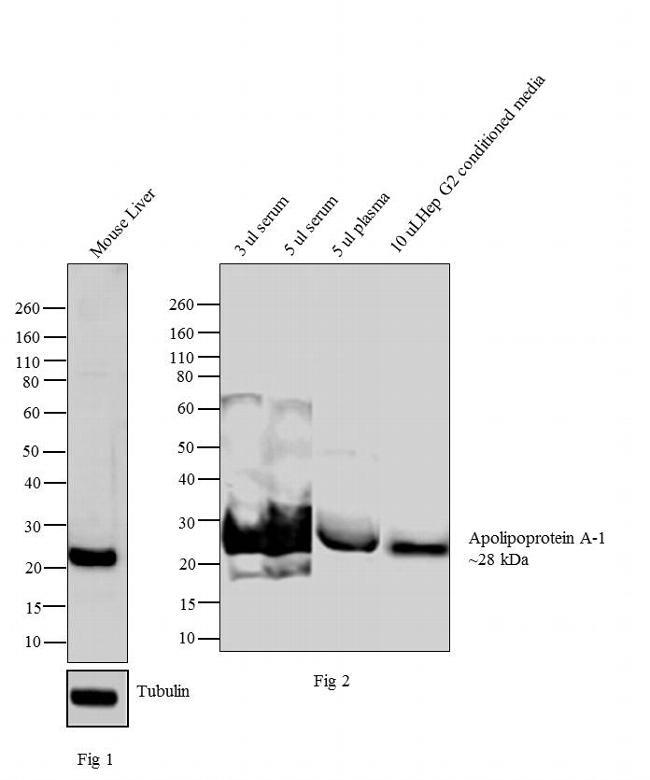
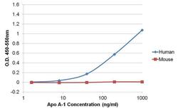
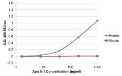
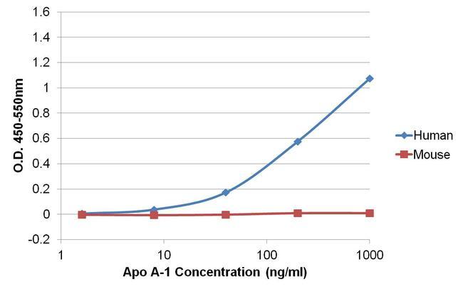
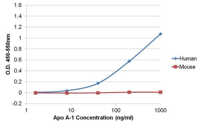
- Sandwich ELISA of Apolipoprotein A-1 was performed by coating wells of a 96-well plate with 100ul of an Apo A-1 rabbit oligoclonal antibody diluted to a concentration of 1 µg/mL in carbonate/bicarbonate buffer overnight at 4C. Wells were blocked with 150ul of StartingBlock T20 (TBS) Blocking Buffer for 30 minutes, and 80ul of recombinant human Apo A-1 or recombinant mouse Apo A-1 was added to the plate at concentrations ranging from 1.6-1000ng/ml and incubated for 1 hour at room temperature. The plate was washed with 1X TBST, and 100ul per well of an Apo A-1 mouse monoclonal antibody was added to each well for 1 hour at room temperature. The plate was washed, and 100ul per well of an HRP-conjugated rabbit anti-mouse IgG cross-adsorbed secondary antibody was incubated for 30 minutes at room temperature. Detection was performed using 1-Step Ultra TMB Substrate, followed by Stop Solution. Absorbances were read on a spectrophotometer at 450-550nm.

- Submitted by
- LSBio (provider)
- Enhanced method
- Genetic validation
- Main image

- Experimental details
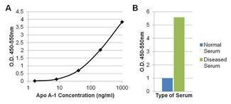
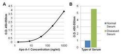
- Sandwich ELISA of Apolipoprotein A-1 was performed by coating wells of a 96-well plate with 100ul of an Apo A-1 rabbit monoclonal antibody at a concentration of 1 µg/mL in carbonate/bicarbonate buffer overnight at 4C. Wells were blocked with 150ul of StartingBlock T20 (TBS) Blocking Buffer for 30 minutes, and 80ul of recombinant human Apo A-1 standards ranging from 1.6-1000ng/ml (A) or 100ul of diluted normal human serum or diluted serum from a patient with dyslipidemia (B) were incubated for 1 hour at room temperature. The plate was washed with 1X TBST, and 100ul per well of an Apo A-1 mouse monoclonal antibody was added to each well for 1 hour at room temperature. The plate was washed, and 100ul per well of a biotinylated rabbit anti-mouse IgG Superclonal secondary antibody was incubated for 1 hour, followed by Streptavidin-HRP for 30 minutes. Detection was performed using 1-Step Ultra TMB Substrate, followed by Stop Solution. Absorbances were read on a spectrophotometer at 450-550nm.
- Submitted by
- LSBio (provider)
- Enhanced method
- Genetic validation
- Main image

- Experimental details
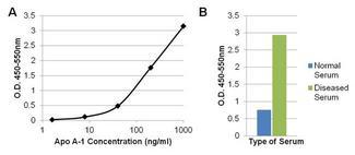
- Sandwich ELISA of Apolipoprotein A-1 was performed by coating wells of a 96-well plate with 100ul of an Apo A-1 rabbit oligoclonal antibody at a concentration of 1 µg/mL in carbonate/bicarbonate buffer overnight at 4C. Wells were blocked with 150ul of StartingBlock T20 (TBS) Blocking Buffer for 30 minutes, and 80ul of recombinant human Apo A-1 standards ranging from 1.6-1000ng/ml (A) or 100ul of diluted normal human serum or diluted serum from a patient with dyslipidemia (B) were incubated for 1 hour at room temperature. The plate was washed with 1X TBST, and 100ul per well of an Apo A-1 mouse monoclonal antibody was added to each well for 1 hour at room temperature. The plate was washed, and 100ul per well of a biotinylated rabbit anti-mouse IgG Superclonal secondary antibody was incubated for 1 hour, followed by Streptavidin-HRP for 30 minutes. Detection was performed using 1-Step Ultra TMB Substrate, followed by Stop Solution. Absorbances were read on a spectrophotometer at 450-550nm.

- Submitted by
- LSBio (provider)
- Main image
- Experimental details
- Sandwich ELISA of Apolipoprotein A-1 was performed by coating wells of a 96-well plate with 100ul of an Apo A-1 rabbit oligoclonal antibody diluted to a concentration of 1 µg/mL in carbonate/bicarbonate buffer overnight at 4C. Wells were blocked with 150ul of StartingBlock T20 (TBS) Blocking Buffer for 30 minutes, and 80ul of recombinant human Apo A-1 or recombinant mouse Apo A-1 was added to the plate at concentrations ranging from 1.6-1000ng/ml and incubated for 1 hour at room temperature. The plate was washed with 1X TBST, and 100ul per well of an Apo A-1 mouse monoclonal antibody was added to each well for 1 hour at room temperature. The plate was washed, and 100ul per well of an HRP-conjugated rabbit anti-mouse IgG cross-adsorbed secondary antibody was incubated for 30 minutes at room temperature. Detection was performed using 1-Step Ultra TMB Substrate, followed by Stop Solution. Absorbances were read on a spectrophotometer at 450-550nm.
- Submitted by
- LSBio (provider)
- Main image

- Experimental details
- Sandwich ELISA of Apolipoprotein A-1 was performed by coating wells of a 96-well plate with 100ul of an Apo A-1 rabbit monoclonal antibody at a concentration of 1 µg/mL in carbonate/bicarbonate buffer overnight at 4C. Wells were blocked with 150ul of StartingBlock T20 (TBS) Blocking Buffer for 30 minutes, and 80ul of recombinant human Apo A-1 standards ranging from 1.6-1000ng/ml (A) or 100ul of diluted normal human serum or diluted serum from a patient with dyslipidemia (B) were incubated for 1 hour at room temperature. The plate was washed with 1X TBST, and 100ul per well of an Apo A-1 mouse monoclonal antibody was added to each well for 1 hour at room temperature. The plate was washed, and 100ul per well of a biotinylated rabbit anti-mouse IgG Superclonal secondary antibody was incubated for 1 hour, followed by Streptavidin-HRP for 30 minutes. Detection was performed using 1-Step Ultra TMB Substrate, followed by Stop Solution. Absorbances were read on a spectrophotometer at 450-550nm.

- Submitted by
- LSBio (provider)
- Main image

- Experimental details
- Sandwich ELISA of Apolipoprotein A-1 was performed by coating wells of a 96-well plate with 100ul of an Apo A-1 rabbit oligoclonal antibody at a concentration of 1 µg/mL in carbonate/bicarbonate buffer overnight at 4C. Wells were blocked with 150ul of StartingBlock T20 (TBS) Blocking Buffer for 30 minutes, and 80ul of recombinant human Apo A-1 standards ranging from 1.6-1000ng/ml (A) or 100ul of diluted normal human serum or diluted serum from a patient with dyslipidemia (B) were incubated for 1 hour at room temperature. The plate was washed with 1X TBST, and 100ul per well of an Apo A-1 mouse monoclonal antibody was added to each well for 1 hour at room temperature. The plate was washed, and 100ul per well of a biotinylated rabbit anti-mouse IgG Superclonal secondary antibody was incubated for 1 hour, followed by Streptavidin-HRP for 30 minutes. Detection was performed using 1-Step Ultra TMB Substrate, followed by Stop Solution. Absorbances were read on a spectrophotometer at 450-550nm.
- Submitted by
- LSBio (provider)
- Main image

- Experimental details
- Sandwich ELISA of Apolipoprotein A-1 was performed by coating wells of a 96-well plate with 100ul of an Apo A-1 rabbit oligoclonal antibody diluted to a concentration of 1 µg/mL in carbonate/bicarbonate buffer overnight at 4C. Wells were blocked with 150ul of StartingBlock T20 (TBS) Blocking Buffer for 30 minutes, and 80ul of recombinant human Apo A-1 or recombinant mouse Apo A-1 was added to the plate at concentrations ranging from 1.6-1000ng/ml and incubated for 1 hour at room temperature. The plate was washed with 1X TBST, and 100ul per well of an Apo A-1 mouse monoclonal antibody was added to each well for 1 hour at room temperature. The plate was washed, and 100ul per well of an HRP-conjugated rabbit anti-mouse IgG cross-adsorbed secondary antibody was incubated for 30 minutes at room temperature. Detection was performed using 1-Step Ultra TMB Substrate, followed by Stop Solution. Absorbances were read on a spectrophotometer at 450-550nm.
- Submitted by
- LSBio (provider)
- Main image
- Experimental details
- Sandwich ELISA of Apolipoprotein A-1 was performed by coating wells of a 96-well plate with 100ul of an Apo A-1 rabbit monoclonal antibody at a concentration of 1 µg/mL in carbonate/bicarbonate buffer overnight at 4C. Wells were blocked with 150ul of StartingBlock T20 (TBS) Blocking Buffer for 30 minutes, and 80ul of recombinant human Apo A-1 standards ranging from 1.6-1000ng/ml (A) or 100ul of diluted normal human serum or diluted serum from a patient with dyslipidemia (B) were incubated for 1 hour at room temperature. The plate was washed with 1X TBST, and 100ul per well of an Apo A-1 mouse monoclonal antibody was added to each well for 1 hour at room temperature. The plate was washed, and 100ul per well of a biotinylated rabbit anti-mouse IgG Superclonal secondary antibody was incubated for 1 hour, followed by Streptavidin-HRP for 30 minutes. Detection was performed using 1-Step Ultra TMB Substrate, followed by Stop Solution. Absorbances were read on a spectrophotometer at 450-550nm.

- Submitted by
- LSBio (provider)
- Main image

- Experimental details
- Sandwich ELISA of Apolipoprotein A-1 was performed by coating wells of a 96-well plate with 100ul of an Apo A-1 rabbit oligoclonal antibody at a concentration of 1 µg/mL in carbonate/bicarbonate buffer overnight at 4C. Wells were blocked with 150ul of StartingBlock T20 (TBS) Blocking Buffer for 30 minutes, and 80ul of recombinant human Apo A-1 standards ranging from 1.6-1000ng/ml (A) or 100ul of diluted normal human serum or diluted serum from a patient with dyslipidemia (B) were incubated for 1 hour at room temperature. The plate was washed with 1X TBST, and 100ul per well of an Apo A-1 mouse monoclonal antibody was added to each well for 1 hour at room temperature. The plate was washed, and 100ul per well of a biotinylated rabbit anti-mouse IgG Superclonal secondary antibody was incubated for 1 hour, followed by Streptavidin-HRP for 30 minutes. Detection was performed using 1-Step Ultra TMB Substrate, followed by Stop Solution. Absorbances were read on a spectrophotometer at 450-550nm.

Supportive validation
- Submitted by
- LSBio (provider)
- Enhanced method
- Genetic validation
- Main image
- Experimental details
- Apolipoprotein A-1 Immunohistochemistry

- Submitted by
- LSBio (provider)
- Enhanced method
- Genetic validation
- Main image

- Experimental details
- Apolipoprotein A-1 Immunohistochemistry