Explore
 Validate
 Learn
Learn Western blot
 ELISA
ELISAAntibody data
- Antibody Data
- Antigen structure
- References [1]
- Comments [0]
- Validations
- Western blot [2]
Submit
Validation data
Reference
Comment
Report error
- Product number
- PA5-19784 - Provider product page

- Provider
- Invitrogen Antibodies
- Product name
- ApoA1 Polyclonal Antibody
- Antibody type
- Polyclonal
- Antigen
- Synthetic peptide
- Description
- This antibody is predicted to react with dog, pig and baboon based on sequence homology.
- Reactivity
- Human
- Host
- Rabbit
- Isotype
- IgG
- Vial size
- 100 µg
- Concentration
- 0.4 mg/mL
- Storage
- Store at 4°C short term. For long term storage, store at -20°C, avoiding freeze/thaw cycles.
Submitted references Inhibition of apolipoprotein B synthesis stimulates endoplasmic reticulum autophagy that prevents steatosis.
Conlon DM, Thomas T, Fedotova T, Hernandez-Ono A, Di Paolo G, Chan RB, Ruggles K, Gibeley S, Liu J, Ginsberg HN
The Journal of clinical investigation 2016 Oct 3;126(10):3852-3867
The Journal of clinical investigation 2016 Oct 3;126(10):3852-3867
No comments: Submit comment
Supportive validation
- Submitted by
- Invitrogen Antibodies (provider)
- Main image

- Experimental details
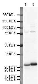
- Western blot analysis of Human Lymph node Tissue Lysate using Product # PA5-19784, Apolipoprotein A I primary antibody at a dilution of 1 µg/mL (lane 1). Staining of Human Testis Tissue Lysate at a dilution of 1 µg/mL (lane 2). Blot treated with a secondary HRP-conjugated Goat polyclonal anti-Rabbit antibody was used at a dilution of 1:3000.
- Submitted by
- Invitrogen Antibodies (provider)
- Main image
- Experimental details
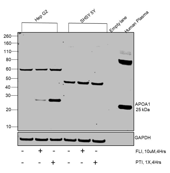
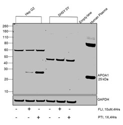
- Western blot was performed using Anti-ApoA1 Polyclonal Antibody(Product # PA5-19784) and a 25kDa band corresponding to ApoA1 was observed across cell lines tested. Whole cell extracts (30 µg lysate) of Hep G2 (Lane 1), Hep G2 treated with FLI, 10 uM, 4 Hrs. (Lane 2), Hep G2 treated with PTI, 1X, 4Hrs. (Lane 3), SH-SY5Y (Lane 4), SH-SY5Y treated with FLI, 10 uM, 4 Hrs (Lane 5), SH-SY5Y treated with PTI, 1X, 4Hrs. (Lane 6), Empty lane (Lane 7) and Human Plasma (Lane 8) were electrophoresed using NuPAGE™ 4-12% Bis-Tris Protein Gel (Product # NP0321BOX). Resolved proteins were then transferred onto a Nitrocellulose membrane (Product # IB23001) by iBlot® 2 Dry Blotting System (Product # IB21001). The blot was probed with the primary antibody (1:1000 Dilution) and detected by chemiluminescence with Goat anti-Mouse IgG (H+L) Superclonal™ Recombinant Secondary Antibody, HRP (Product # A28177, 1:4000 dilution) using the iBright FL 1000 (Product # A32752). Chemiluminescent detection was performed using Novex® ECL Chemiluminescent Substrate Reagent Kit (Product # WP20005). Expression of APOA1 was found to be higher in Hep G2 cell line as compared to SH-SY 5Y.