Explore
 Validate
 Learn
Learn Immunocytochemistry
 Immunohistochemistry
Antibody data
- Antibody Data
- Antigen structure
- References [11]
- Comments [0]
- Validations
- Immunocytochemistry [1]
Submit
Validation data
Reference
Comment
Report error
- Product number
- HPA037606 - Provider product page

- Provider
- Atlas Antibodies
- Proper citation
- Atlas Antibodies Cat#HPA037606, RRID:AB_10672498
- Product name
- Anti-CEP164
- Antibody type
- Polyclonal
- Description
- Polyclonal Antibody against Human CEP164, Gene description: centrosomal protein 164kDa, Alternative Gene Names: KIAA1052, NPHP15, Validated applications: IHC, ICC, Uniprot ID: Q9UPV0, Storage: Store at +4°C for short term storage. Long time storage is recommended at -20°C.
- Reactivity
- Human
- Host
- Rabbit
- Conjugate
- Unconjugated
- Isotype
- IgG
- Vial size
- 100 µl
- Concentration
- 0.2 mg/ml
- Storage
- Store at +4°C for short term storage. Long time storage is recommended at -20°C.
- Handling
- The antibody solution should be gently mixed before use.
Submitted references Deup1 Expression Interferes with Multiciliated Differentiation.
3D-Structured Illumination Microscopy of Centrosomes in Human Cell Lines
CEP78 functions downstream of CEP350 to control biogenesis of primary cilia by negatively regulating CP110 levels
The molecular dynamics of subdistal appendages in multi-ciliated cells.
Centrosome anchoring regulates progenitor properties and cortical formation
A liquid-like spindle domain promotes acentrosomal spindle assembly in mammalian oocytes
Centrobin controls primary ciliogenesis in vertebrates
KIF13B establishes a CAV1-enriched microdomain at the ciliary transition zone to promote Sonic hedgehog signalling
Mutations in KIAA0586 Cause Lethal Ciliopathies Ranging from a Hydrolethalus Phenotype to Short-Rib Polydactyly Syndrome
DCDC2 mutations cause a renal-hepatic ciliopathy by disrupting Wnt signaling.
Mutations of CEP83 Cause Infantile Nephronophthisis and Intellectual Disability
Shin M, Lee J, Lee H, Kumar V, Kim J, Park S
Molecules and cells 2023 Dec 31;46(12):746-756
Molecules and cells 2023 Dec 31;46(12):746-756
3D-Structured Illumination Microscopy of Centrosomes in Human Cell Lines
Frikstad K, Schink K, Gilani S, Pedersen L, Patzke S
BIO-PROTOCOL 2022;12(6)
BIO-PROTOCOL 2022;12(6)
CEP78 functions downstream of CEP350 to control biogenesis of primary cilia by negatively regulating CP110 levels
Gonçalves A, Hasselbalch S, Joensen B, Patzke S, Martens P, Ohlsen S, Quinodoz M, Nikopoulos K, Suleiman R, Damsø Jeppesen M, Weiss C, Christensen S, Rivolta C, Andersen J, Farinelli P, Pedersen L
eLife 2021;10
eLife 2021;10
The molecular dynamics of subdistal appendages in multi-ciliated cells.
Ryu H, Lee H, Lee J, Noh H, Shin M, Kumar V, Hong S, Kim J, Park S
Nature communications 2021 Jan 27;12(1):612
Nature communications 2021 Jan 27;12(1):612
Centrosome anchoring regulates progenitor properties and cortical formation
Shao W, Yang J, He M, Yu X, Lee C, Yang Z, Joyner A, Anderson K, Zhang J, Tsou M, Shi H, Shi S
Nature 2020;580(7801):106-112
Nature 2020;580(7801):106-112
A liquid-like spindle domain promotes acentrosomal spindle assembly in mammalian oocytes
So C, Seres K, Steyer A, Mönnich E, Clift D, Pejkovska A, Möbius W, Schuh M
Science 2019;364(6447)
Science 2019;364(6447)
Centrobin controls primary ciliogenesis in vertebrates
Ogungbenro Y, Tena T, Gaboriau D, Lalor P, Dockery P, Philipp M, Morrison C
Journal of Cell Biology 2018;217(4):1205-1215
Journal of Cell Biology 2018;217(4):1205-1215
KIF13B establishes a CAV1-enriched microdomain at the ciliary transition zone to promote Sonic hedgehog signalling
Schou K, Mogensen J, Morthorst S, Nielsen B, Aleliunaite A, Serra-Marques A, Fürstenberg N, Saunier S, Bizet A, Veland I, Akhmanova A, Christensen S, Pedersen L
Nature Communications 2017;8(1)
Nature Communications 2017;8(1)
Mutations in KIAA0586 Cause Lethal Ciliopathies Ranging from a Hydrolethalus Phenotype to Short-Rib Polydactyly Syndrome
Alby C, Piquand K, Huber C, Megarbané A, Ichkou A, Legendre M, Pelluard F, Encha-Ravazi F, Abi-Tayeh G, Bessières B, El Chehadeh-Djebbar S, Laurent N, Faivre L, Sztriha L, Zombor M, Szabó H, Failler M, Garfa-Traore M, Bole C, Nitschké P, Nizon M, Elkhartoufi N, Clerget-Darpoux F, Munnich A, Lyonnet S, Vekemans M, Saunier S, Cormier-Daire V, Attié-Bitach T, Thomas S
The American Journal of Human Genetics 2015;97(2):311-318
The American Journal of Human Genetics 2015;97(2):311-318
DCDC2 mutations cause a renal-hepatic ciliopathy by disrupting Wnt signaling.
Schueler M, Braun DA, Chandrasekar G, Gee HY, Klasson TD, Halbritter J, Bieder A, Porath JD, Airik R, Zhou W, LoTurco JJ, Che A, Otto EA, Böckenhauer D, Sebire NJ, Honzik T, Harris PC, Koon SJ, Gunay-Aygun M, Saunier S, Zerres K, Bruechle NO, Drenth JP, Pelletier L, Tapia-Páez I, Lifton RP, Giles RH, Kere J, Hildebrandt F
American journal of human genetics 2015 Jan 8;96(1):81-92
American journal of human genetics 2015 Jan 8;96(1):81-92
Mutations of CEP83 Cause Infantile Nephronophthisis and Intellectual Disability
Failler M, Gee H, Krug P, Joo K, Halbritter J, Belkacem L, Filhol E, Porath J, Braun D, Schueler M, Frigo A, Alibeu O, Masson C, Brochard K, Hurault de Ligny B, Novo R, Pietrement C, Kayserili H, Salomon R, Gubler M, Otto E, Antignac C, Kim J, Benmerah A, Hildebrandt F, Saunier S
The American Journal of Human Genetics 2014;94(6):905-914
The American Journal of Human Genetics 2014;94(6):905-914
No comments: Submit comment
Supportive validation
- Submitted by
- Atlas Antibodies (provider)
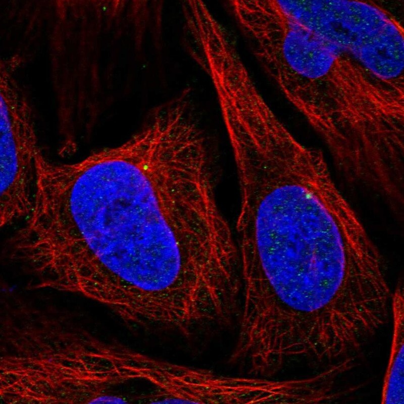
- Main image

- Experimental details
- Immunofluorescent staining of human cell line U-2 OS shows localization to centrosome.
- Sample type
- Human