 Explore
 Validate
 Learn
Learn Western blot
Antibody data
- Antibody Data
- Antigen structure
- References [0]
- Comments [0]
- Validations
- Western blot [2]
- Immunocytochemistry [2]
- Immunohistochemistry [2]
- Flow cytometry [2]
Submit
Validation data
Reference
Comment
Report error
- Product number
- PA5-35370 - Provider product page

- Provider
- Invitrogen Antibodies
- Product name
- OAT Polyclonal Antibody
- Antibody type
- Polyclonal
- Antigen
- Synthetic peptide
- Reactivity
- Human, Mouse, Rat
- Host
- Rabbit
- Isotype
- IgG
- Vial size
- 400 μL
- Concentration
- 0.5 mg/mL
- Storage
- Store at 4°C short term. For long term storage, store at -20°C, avoiding freeze/thaw cycles.
No comments: Submit comment
Supportive validation
- Submitted by
- Invitrogen Antibodies (provider)
- Main image

- Experimental details
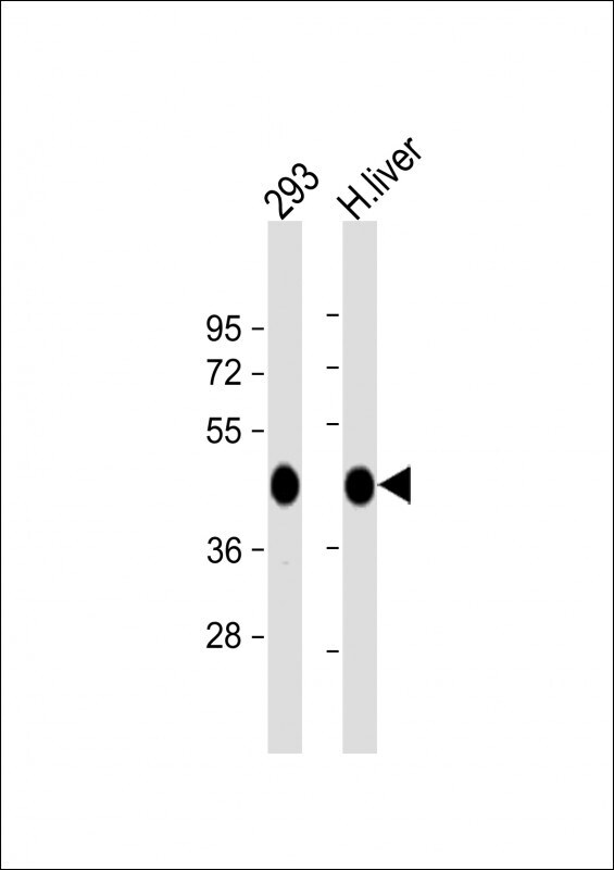
- Western blot analysis of OAT in various lysates. Samples were incubated with OAT polyclonal antibody (Product # PA5-35370) using a dilution of 1:1,000 followed by Goat Anti-Rabbit IgG, (H+L), Peroxidase conjugated at a dilution of 1:10,000. Lysates/proteins: 20 µg per lane. Lane 1: 293 whole cell lysate; Lane 2: human liver lysate. Predicted band size: 49 kDa. Blocking/Dilution buffer: 5% NFDM/TBST.
- Submitted by
- Invitrogen Antibodies (provider)
- Main image
- Experimental details
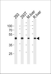
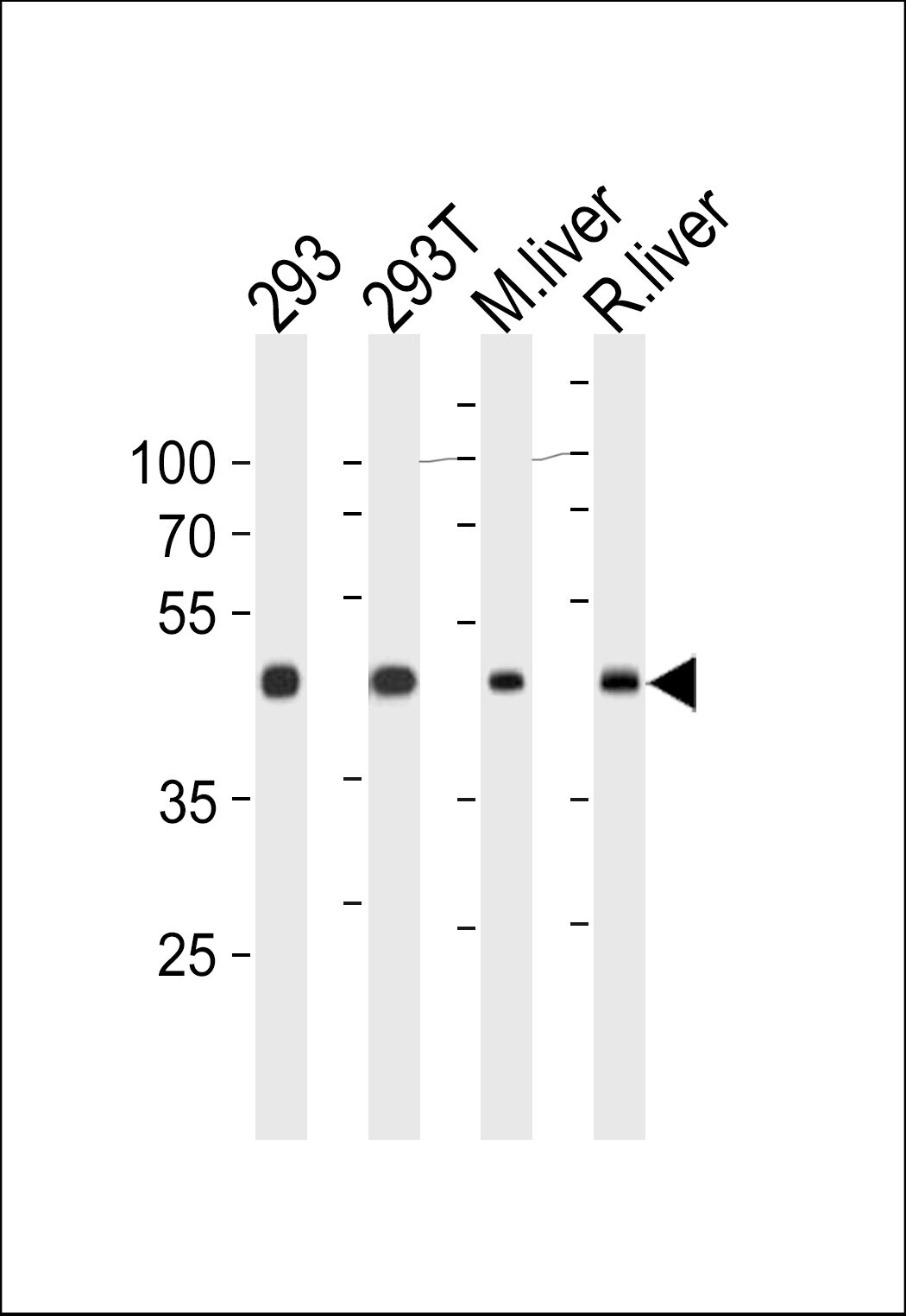
- Western blot analysis of OAT in 293, 293T cell line, mouse liver and rat liver tissue lysates. Samples were incubated with OAT polyclonal antibody (Product # PA5-35370). Lysates: 35 µg/lane. OAT protein (arrow).
Supportive validation
- Submitted by
- Invitrogen Antibodies (provider)
- Main image
- Experimental details
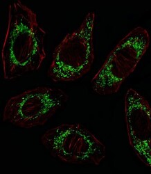
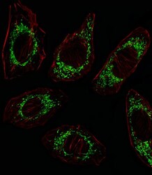
- Immunofluorescent analysis of OAT in mitochondria of A549 cells using an OAT polyclonal antibody (Product # PA5-35370) followed by detection using a fluorescent conjugated secondary antibody (green). Cytoplasmic actin was stained with a fluorescent red phalloidin (7units/mL, 1 h at 37ºC).

- Submitted by
- Invitrogen Antibodies (provider)
- Main image
- Experimental details
- Immunocytochemistry analysis of OAT in A549 cells. Samples were incubated with OAT polyclonal antibody (Product # PA5-35370) using a dilution of 1:25 for 1 h at 37°C followed by Alexa Fluor® 488 conjugated donkey anti-rabbit antibody (green) at a dilution of 1:400 for 50 min at 37°C. Cells were fixed with 4% PFA (20 min) and permeabilized with Triton X-100 (0.1%, 10 min). Cytoplasmic actin was counterstained with Alexa Fluor® 555 (red) conjugated Phalloidin (7 units/mL, 1 h at 37°C). OAT immunoreactivity is localized to Mitochondrion significantly.

Supportive validation
- Submitted by
- Invitrogen Antibodies (provider)
- Main image
- Experimental details
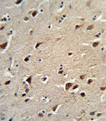
- Immunohistochemistry analysis of OAT in formalin-fixed and paraffin-embedded human brain tissue. Samples were incubated with OAT polyclonal antibody (Product # PA5-35370) which was peroxidase-conjugated to the secondary antibody, followed by DAB staining. This data demonstrates the use of this antibody for immunohistochemistry; clinical relevance has not been evaluated.
- Submitted by
- Invitrogen Antibodies (provider)
- Main image

- Experimental details
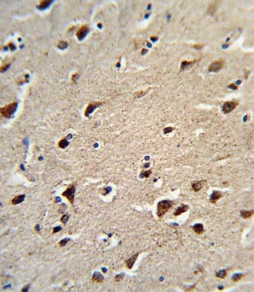
- Immunohistochemistry analysis of OAT in formalin-fixed and paraffin-embedded human brain tissue. Samples were incubated with OAT polyclonal antibody (Product # PA5-35370) which was peroxidase-conjugated to the secondary antibody, followed by DAB staining. This data demonstrates the use of this antibody for immunohistochemistry; clinical relevance has not been evaluated.

Supportive validation
- Submitted by
- Invitrogen Antibodies (provider)
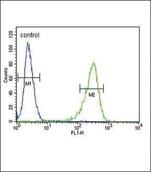
- Main image
- Experimental details
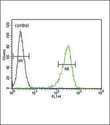
- Flow cytometry analysis of OAT in 293 cells (right) compared to a negative control (left) using an OAT polyclonal antibody (Product # PA5-35370) followed by detection using a FITC-conjugated goat-anti-rabbit secondary antibody.
- Submitted by
- Invitrogen Antibodies (provider)
- Main image
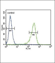
- Experimental details
- Flow cytometry of OAT in 293 cells (right histogram). Samples were incubated with OAT polyclonal antibody (Product # PA5-35370) followed by FITC-conjugated goat-anti-rabbit secondary antibody. Negative control cell (left histogram).
