Explore
 Validate
 Learn
Learn Western blot
Antibody data
- Antibody Data
- Antigen structure
- References [0]
- Comments [0]
- Validations
- Western blot [1]
- Immunocytochemistry [1]
- Immunohistochemistry [1]
- Flow cytometry [1]
Submit
Validation data
Reference
Comment
Report error
- Product number
- TA325081 - Provider product page

- Provider
- OriGene
- Product name
- Rabbit polyclonal OAT Antibody (N-term)
- Antibody type
- Polyclonal
- Description
- Rabbit polyclonal OAT Antibody (N-term)
- Host
- Rabbit
- Conjugate
- Unconjugated
- Epitope
- OAT
- Isotype
- IgG
- Antibody clone number
- NULL
- Vial size
- 400 µl
- Concentration
- 0.5 mg/ml
No comments: Submit comment
Supportive validation
- Submitted by
- OriGene (provider)
- Main image
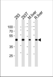
- Experimental details
- OAT Antibody (N-term) (Cat. #TA325081) western blot analysis in 293,293T cell line ,mouse liver and rat liver tissue lysates (35ug/lane).This demonstrates the OAT antibody detected the OAT protein (arrow).
- Validation comment
- WB
Supportive validation
- Submitted by
- OriGene (provider)
- Main image

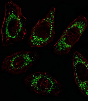
- Experimental details
- IF image of A549 cell stained with OAT Antibody (N-term)(Cat#TA325081/SA100310AG).A549 cells were incubated with OAT primary antibody (1:25, 1 h at 37?). For secondary antibody, Alexa Fluor? 488 conjugated donkey anti-rabbit antibody (green) was used (1:400).Cytoplasmic actin was counterstained with Alexa Fluor? 555 (red) conjugated Phalloidin (7 units/ml).OAT immunoreactivity is localized to Mitochondrion significantly.
- Validation comment
- IF
Supportive validation
- Submitted by
- OriGene (provider)
- Main image
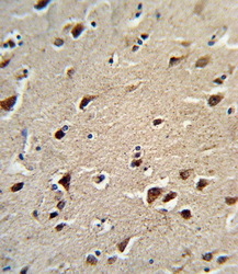
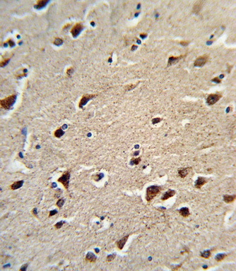
- Experimental details
- Formalin-fixed and paraffin-embedded human brain tissue reacted with OAT Antibody (N-term), which was peroxidase-conjugated to the secondary antibody, followed by DAB staining. This data demonstrates the use of this antibody for immunohistochemistry; clinical relevance has not been evaluated.
- Validation comment
- IHC
Supportive validation
- Submitted by
- OriGene (provider)
- Main image
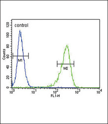
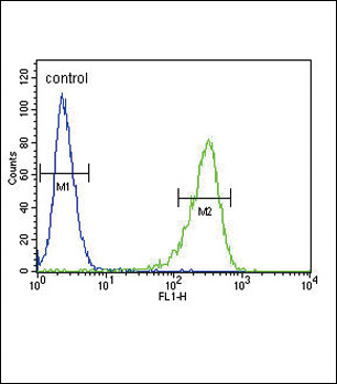
- Experimental details
- OAT Antibody (N-term) (Cat. #TA325081) flow cytometric analysis of 293 cells (right histogram) compared to a negative control cell (left histogram).FITC-conjugated goat-anti-rabbit secondary antibodies were used for the analysis.
- Validation comment
- FC