Explore
 Validate
 Learn
Learn Western blot
 Immunocytochemistry
 Immunoprecipitation
Antibody data
- Antibody Data
- Antigen structure
- References [1]
- Comments [0]
- Validations
- Immunocytochemistry [5]
- Immunohistochemistry [3]
- Other assay [2]
Submit
Validation data
Reference
Comment
Report error
- Product number
- PA5-34939 - Provider product page

- Provider
- Invitrogen Antibodies
- Product name
- hnRNP A2B1 Polyclonal Antibody
- Antibody type
- Polyclonal
- Antigen
- Recombinant full-length protein
- Description
- Recommended positive controls: 293T, A431, HeLa, HepG2, Neuro2A, C8D30, NIH-3T3, Raw264.7, C2C12, PC-12. Predicted reactivity: Mouse (100%), Rat (100%), Rhesus Monkey (100%), Bovine (100%). Store product as a concentrated solution. Centrifuge briefly prior to opening the vial.
- Reactivity
- Human, Mouse, Rat
- Host
- Rabbit
- Isotype
- IgG
- Vial size
- 100 μL
- Concentration
- 1.09 mg/mL
- Storage
- Store at 4°C short term. For long term storage, store at -20°C, avoiding freeze/thaw cycles.
Submitted references YBX-1 mediated sorting of miR-133 into hypoxia/reoxygenation-induced EPC-derived exosomes to increase fibroblast angiogenesis and MEndoT.
Lin F, Zeng Z, Song Y, Li L, Wu Z, Zhang X, Li Z, Ke X, Hu X
Stem cell research & therapy 2019 Aug 23;10(1):263
Stem cell research & therapy 2019 Aug 23;10(1):263
No comments: Submit comment
Supportive validation
- Submitted by
- Invitrogen Antibodies (provider)
- Main image

- Experimental details
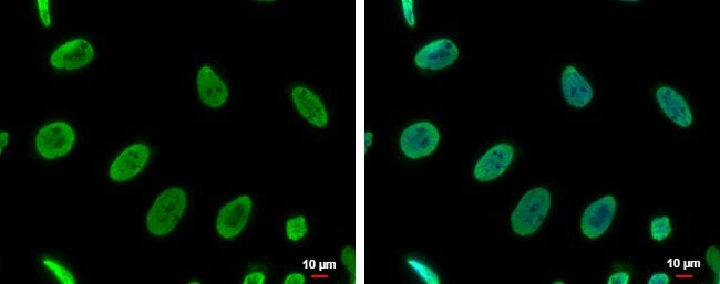
- Immunofluorescent analysis of hnRNP A2B1 showing staining in the nucleus of HeLa cells. HeLa cells were fixed in 4% paraformaldehyde at RT for 15 min and stained using a hnRNP A2B1 polyclonal antibody (Product # PA5-34939) diluted at 1:500. Blue: Hoechst 33342 staining. Scale bar = 10µm.
- Submitted by
- Invitrogen Antibodies (provider)
- Main image
- Experimental details
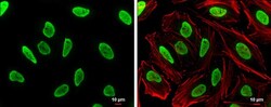
- Immunocytochemistry-Immunofluorescence analysis of hnRNP A2B1 was performed in HeLa cells fixed in 4% paraformaldehyde at RT for 15 min. Green: hnRNP A2B1 Polyclonal Antibody (Product # PA5 34939) diluted at 1:500. Red: phalloidin, a cytoskeleton marker. Scale bar = 10 µm.

- Submitted by
- Invitrogen Antibodies (provider)
- Main image

- Experimental details
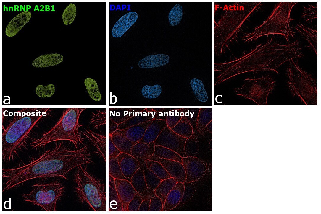
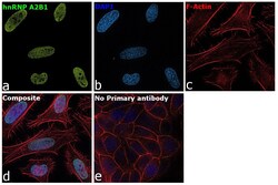
- Immunofluorescence analysis of hnRNP A2B1 was performed using 70% confluent log phase MCF7 cells. The cells were fixed with 4% paraformaldehyde for 10 minutes, permeabilized with 0.1% Triton™ X-100 for 15 minutes, and blocked with 2% BSA for 45 minutes at room temperature. The cells were labeled with hnRNP A2B1 Polyclonal Antibody (Product # PA5-34939) at 1:200 in 0.1% BSA, incubated at 4 degree celsius overnight and then labeled with Donkey anti-Rabbit IgG (H+L) Highly Cross-Adsorbed Secondary Antibody, Alexa Fluor Plus 488 (Product # A32790), (1:2000), for 45 minutes at room temperature (Panel a: Green). Nuclei (Panel b:Blue) were stained with ProLong™ Diamond Antifade Mountant with DAPI (Product # P36962). F-actin (Panel c: Red) was stained with Rhodamine Phalloidin (Product # R415, 1:300). Panel d represents the merged image showing Nuclear localization. Panel e represents control cells with no primary antibody to assess background. The images were captured at 60X magnification.
- Submitted by
- Invitrogen Antibodies (provider)
- Main image

- Experimental details
- Immunocytochemistry-Immunofluorescence analysis of hnRNP A2B1 was performed in HeLa cells fixed in 4% paraformaldehyde at RT for 15 min. Green: hnRNP A2B1 Polyclonal Antibody (Product # PA5 34939) diluted at 1:500. Red: phalloidin, a cytoskeleton marker. Scale bar = 10 µm.

- Submitted by
- Invitrogen Antibodies (provider)
- Main image
- Experimental details
- Immunofluorescence analysis of hnRNP A2B1 was performed using 70% confluent log phase MCF7 cells. The cells were fixed with 4% paraformaldehyde for 10 minutes, permeabilized with 0.1% Triton™ X-100 for 15 minutes, and blocked with 2% BSA for 45 minutes at room temperature. The cells were labeled with hnRNP A2B1 Polyclonal Antibody (Product # PA5-34939) at 1:200 in 0.1% BSA, incubated at 4 degree celsius overnight and then labeled with Donkey anti-Rabbit IgG (H+L) Highly Cross-Adsorbed Secondary Antibody, Alexa Fluor Plus 488 (Product # A32790), (1:2000), for 45 minutes at room temperature (Panel a: Green). Nuclei (Panel b:Blue) were stained with ProLong™ Diamond Antifade Mountant with DAPI (Product # P36962). F-actin (Panel c: Red) was stained with Rhodamine Phalloidin (Product # R415, 1:300). Panel d represents the merged image showing Nuclear localization. Panel e represents control cells with no primary antibody to assess background. The images were captured at 60X magnification.

Supportive validation
- Submitted by
- Invitrogen Antibodies (provider)
- Main image
- Experimental details
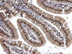
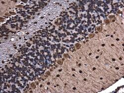
- hnRNP A2B1 Polyclonal Antibody detects hnRNP A2B1 protein at cytosol and nucleus on mouse duodenum by immunohistochemical analysis. Sample: Paraffin-embedded mouse duodenum. HnRNP A2B1 Polyclonal Antibody (Product # PA5-34939) dilution: 1:500. Antigen Retrieval: EDTA based buffer, pH 8.0, 15 min.
- Submitted by
- Invitrogen Antibodies (provider)
- Main image
- Experimental details
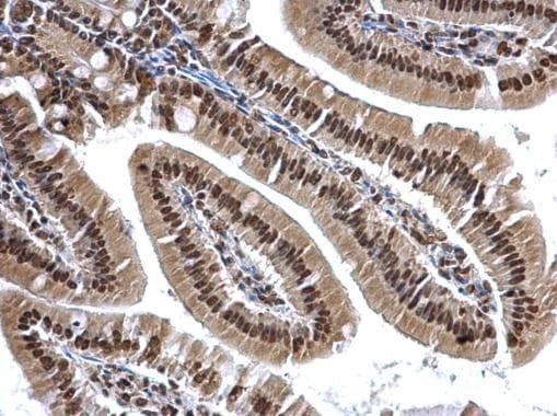
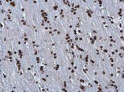
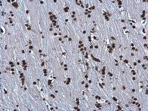
- Immunohistochemistry (Paraffin) analysis of hnRNP A2B1 was performed in paraffin-embedded mouse brain tissue using hnRNP A2B1 Polyclonal Antibody (Product # PA5-34939) at a dilution of 1:500.
- Submitted by
- Invitrogen Antibodies (provider)
- Main image
- Experimental details
- Immunohistochemistry (Paraffin) analysis of hnRNP A2B1 was performed in paraffin-embedded mouse brain tissue using hnRNP A2B1 Polyclonal Antibody (Product # PA5-34939) at a dilution of 1:500.

Supportive validation
- Submitted by
- Invitrogen Antibodies (provider)
- Main image
- Experimental details
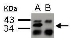
- hnRNPA2B1 antibody immunoprecipitates hnRNPA2B1 protein in IP experiments. IP Sample: 4500 µg A549 whole cell lysate/extract A. Immunoprecipitation of hnRNPA2B1 protein by 2 µg of hnRNPA2B1 antibody (Product # PA5-34939) B. Control with 2 µg of preimmune rabbit IgG The immunoprecipitated hnRNPA2B1 protein was detected by hnRNPA2B1 antibody diluted at 1:2,000.
- Submitted by
- Invitrogen Antibodies (provider)
- Main image

- Experimental details
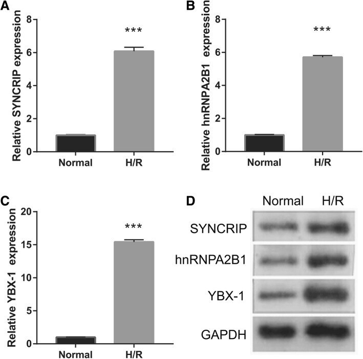
- Fig. 6 Measurements of YBX-1, SYNCRIP, and hnRNPA2B1 in EPCs by qRT-PCR ( a - c ) and western blotting ( d ) after 48-h H/R treatment