 Explore
 Validate
 Learn
Learn Western blot
 Immunoprecipitation
Antibody data
- Antibody Data
- Antigen structure
- References [0]
- Comments [0]
- Validations
- Western blot [1]
- Immunocytochemistry [2]
Submit
Validation data
Reference
Comment
Report error
- Product number
- PA5-19577 - Provider product page

- Provider
- Invitrogen Antibodies
- Product name
- hnRNP A2B1 Polyclonal Antibody
- Antibody type
- Polyclonal
- Antigen
- Synthetic peptide
- Description
- Heat mediated antigen retrieval recommended prior to tissue staining. This antibody is predicted to react with chicken, dog and Xenopus laevis based on sequence homology.
- Reactivity
- Human, Mouse, Rat
- Host
- Rabbit
- Isotype
- IgG
- Vial size
- 100 µg
- Concentration
- 1 mg/mL
- Storage
- Store at 4°C short term. For long term storage, store at -20°C, avoiding freeze/thaw cycles.
No comments: Submit comment
Supportive validation
- Submitted by
- Invitrogen Antibodies (provider)
- Main image

- Experimental details
- Western blot analysis of HeLa Whole Cell Lysate using Product # PA5-19577, hnRNP A2B1 primary antibody at a dilution of 1 µg/mL. Blot treated with a secondary IR Dye680-conjugated Goat polyclonal anti-Rabbit antibody was used at a dilution of 1:10000.

Supportive validation
- Submitted by
- Invitrogen Antibodies (provider)
- Main image
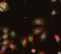
- Experimental details
- Immunofluorescent staining of MCF7 cells using Product # PA5-19577, anti-hnRNP A2B1 antibody. The cells were fixed with PFA (4%)for 10 minutes, permabilised PBS-T for 20 minutes and exposed to the primary antibody at a concentration of 5 µg/mL for 1 hour at room temp. A solution of BSA (1%), normal goat serum (10%) and glycine (0.3 M) was used to quench autofluorescence and block non-specific protein-protein interactions. The secondary antibody was a 448 fluorescence conjugated Goat anti-rabbit IgG (green) at a dilution of 1:1000. A WGA- 594 fluorescent conjugated stain was used to label plasma membranes (red) and the nuclei stain was DAPI (blue).
