 Explore
 Validate
 Learn
Learn Western blot
 Immunohistochemistry
Antibody data
- Antibody Data
- Antigen structure
- References [0]
- Comments [0]
- Validations
- Immunohistochemistry [1]
Submit
Validation data
Reference
Comment
Report error
- Product number
- PA5-26631 - Provider product page

- Provider
- Invitrogen Antibodies
- Product name
- AANAT Polyclonal Antibody
- Antibody type
- Polyclonal
- Antigen
- Synthetic peptide
- Description
- PA5-26631 has successfully been used to detect serotonin N-AT in both WB and IF applications.
- Reactivity
- Human
- Host
- Rabbit
- Isotype
- IgG
- Vial size
- 400 μL
- Concentration
- 0.5 mg/mL
- Storage
- Store at 4°C short term. For long term storage, store at -20°C, avoiding freeze/thaw cycles.
No comments: Submit comment
Supportive validation
- Submitted by
- Invitrogen Antibodies (provider)
- Main image
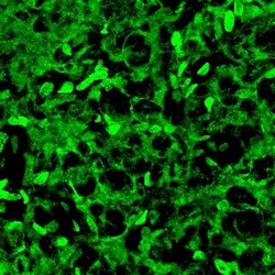
- Experimental details
- Immunofluorescence was performed on sections of adult rat pineal. Tissue sections on slides were probed for 24 h at 4°C in a humidified chamber with rabbit anti-AANAT (Product # PA5-26631) at an antibody concentration of 1-2 µg/mL diluted in 50 mM Tris-HCl, pH 7.4, containing 1.5% NaCl, 0.3% Triton X-100 (TBST) and 4% normal goat serum. After overnight incubation, sections were washed extensively for 1 h in TBST, and detection of primary antibody was performed for 1.5 h at room temperature with AlexaFluor-488-conjugated donkey anti-rabbit diluted 1:600 in TBST. Sections were then washed in TBST, then in TBS (without triton) and then coversliped with anti-fade medium. Images were taken on a Zeiss Z2 scanning microscope at x40 objective magnification, and show immunofluorescence labelling of AANAT localized to pinealocytes. Data courtesy of Dr. James Nagy's laboratory.
