 Explore
 Validate
 Learn
Learn Western blot
Antibody data
- Antibody Data
- Antigen structure
- References [0]
- Comments [0]
- Validations
- Western blot [1]
- Immunocytochemistry [2]
- Immunohistochemistry [1]
- Flow cytometry [1]
Submit
Validation data
Reference
Comment
Report error
- Product number
- AGR-031-200UL - Provider product page

- Provider
- Invitrogen Antibodies
- Product name
- Ghrelin Receptor (GHSR) (extracellular) Polyclonal Antibody
- Antibody type
- Polyclonal
- Antigen
- Other
- Reactivity
- Human, Mouse, Rat
- Host
- Rabbit
- Isotype
- IgG
- Vial size
- 200 µL
- Concentration
- 1 mg/mL
- Storage
- -20° C, Avoid Freeze/Thaw Cycles
No comments: Submit comment
Supportive validation
- Submitted by
- Invitrogen Antibodies (provider)
- Main image

- Experimental details
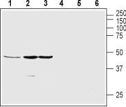
- Western blot analysis of rat pancreas (lanes 1 and 4), human SH-SY5Y neuroblastoma (lanes 2 and 5) and mouse MS1 endothelial (lanes 3 and 6) cells line lysates: - 1-3. Anti-Ghrelin Receptor (GHSR) (extracellular) Antibody (#AGR-031), (1:200).4-6. Anti-Ghrelin Receptor (GHSR) (extracellular) Antibody , preincubated with Ghrelin Receptor/GHSR (extracellular) Blocking Peptide (#BLP-GR031).
Supportive validation
- Submitted by
- Invitrogen Antibodies (provider)
- Main image
- Experimental details
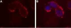
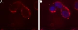
- Expression of Ghrelin receptor in human SH-SY5Y cell line - Cell surface detection of Ghrelin receptor in intact living human neuroblastoma (SH-SY5Y) cells. A. Extracellular staining of cells with Anti-Ghrelin Receptor (GHSR) (extracellular) Antibody (#AGR-031), (red), (1:50) followed by goat Anti-rabbit-AlexaFluor-594 secondary Antibody . B. DAPI is used as the counterstain (blue) merged with Anti-Ghrelin Receptor Antibody staining.
- Submitted by
- Invitrogen Antibodies (provider)
- Main image
- Experimental details
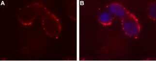
- Expression of Ghrelin receptor in human SH-SY5Y cell line - Cell surface detection of Ghrelin receptor in intact living human neuroblastoma (SH-SY5Y) cells. A. Extracellular staining of cells with Anti-Ghrelin Receptor (GHSR) (extracellular) Antibody (#AGR-031), (red), (1:50) followed by goat Anti-rabbit-AlexaFluor-594 secondary Antibody . B. DAPI is used as the counterstain (blue) merged with Anti-Ghrelin Receptor Antibody staining.
Supportive validation
- Submitted by
- Invitrogen Antibodies (provider)
- Main image
- Experimental details
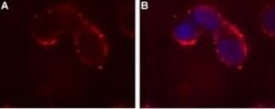
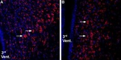
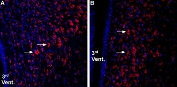
- Expression of Ghrelin receptor in rat and mouse hypothalamus - Immunohistochemical staining of rat and mouse hypothalamic paraventricular nucleus (PVN) using Anti-Ghrelin Receptor (GHSR) (extracellular) Antibody (#AGR-031), (1:800). A. Ghrelin receptor 1 staining (red) in mouse appears in cells of the PVN (arrows). B. Ghrelin receptor staining in rat is similar. Nuclear staining using DAPI as the counterstain (blue).
Supportive validation
- Submitted by
- Invitrogen Antibodies (provider)
- Main image
- Experimental details
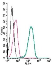
- Cell surface detection of Ghrelin Receptor by indirect flow cytometry in live intact human THP-1 monocytic leukemia cells: - (black line) cells. (red) Cells + goat- Anti-rabbit-FITC. (green) Cells + Anti-Ghrelin Receptor (GHSR) (extracellular) Antibody (#AGR-031), (2.5μg) + goat- Anti-rabbit-FITC.
