Explore
 Validate
 Learn
Learn Western blot
 Immunocytochemistry
Antibody data
- Antibody Data
- Antigen structure
- References [2]
- Comments [0]
- Validations
- Immunocytochemistry [2]
- Immunohistochemistry [1]
- Other assay [1]
Submit
Validation data
Reference
Comment
Report error
- Product number
- PA5-28752 - Provider product page

- Provider
- Invitrogen Antibodies
- Product name
- GHSR Polyclonal Antibody
- Antibody type
- Polyclonal
- Antigen
- Synthetic peptide
- Description
- Recommended positive controls: NT2D1, PC-3, U87-MG, SK-N-SH, mouse hypothalamus, mouse brain, rat brain. Store product as a concentrated solution. Centrifuge briefly prior to opening the vial.
- Reactivity
- Human, Mouse
- Host
- Rabbit
- Isotype
- IgG
- Vial size
- 100 μL
- Concentration
- 1 mg/mL
- Storage
- Store at 4°C short term. For long term storage, store at -20°C, avoiding freeze/thaw cycles.
Submitted references Defective dystrophic thymus determines degenerative changes in skeletal muscle.
ASSOCIATION OF GHRELIN RECEPTOR AND INFLAMMATION IN PERI-ATRIAL ADIPOSE TISSUE FROM OBESE PATIENTS WITH POSTOPERATIVE ATRIAL FIBRILLATION.
Farini A, Sitzia C, Villa C, Cassani B, Tripodi L, Legato M, Belicchi M, Bella P, Lonati C, Gatti S, Cerletti M, Torrente Y
Nature communications 2021 Apr 8;12(1):2099
Nature communications 2021 Apr 8;12(1):2099
ASSOCIATION OF GHRELIN RECEPTOR AND INFLAMMATION IN PERI-ATRIAL ADIPOSE TISSUE FROM OBESE PATIENTS WITH POSTOPERATIVE ATRIAL FIBRILLATION.
Mocanu V, Timofte D, Oboroceanu T, Cretu-Silivestru IS, Pricope-Veselin A, Moraru M, Butcovan D
Acta endocrinologica (Bucharest, Romania : 2005) 2020 Jul-Sep;16(3):298-302
Acta endocrinologica (Bucharest, Romania : 2005) 2020 Jul-Sep;16(3):298-302
No comments: Submit comment
Supportive validation
- Submitted by
- Invitrogen Antibodies (provider)
- Main image

- Experimental details
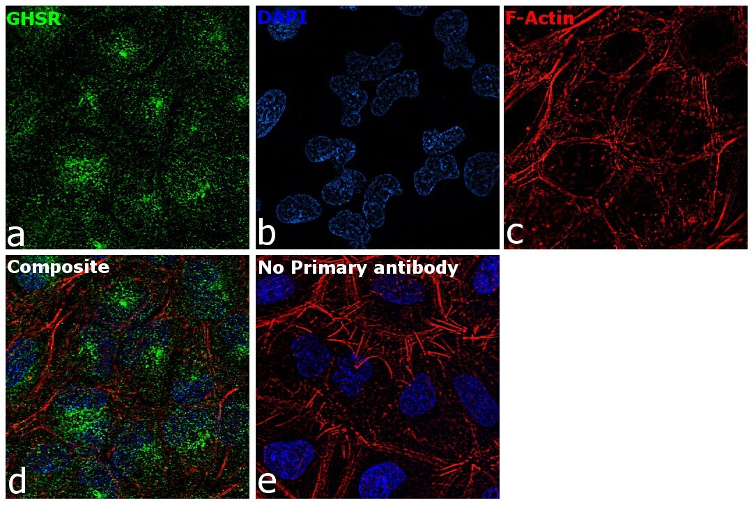
- Immunofluorescence analysis of GHSR was performed using 70% confluent log phase HeLa cells. The cells were fixed with 4% paraformaldehyde for 10 minutes, permeabilized with 0.1% Triton™ X-100 for 15 minutes, and blocked with 1% BSA for 1 hour at room temperature. The cells were labeled with GHSR Polyclonal Antibody (Product # PA5-28752) at 5 microgram/mL in 0.1% BSA, incubated at 4 degree Celsius overnight and then labeled with Goat anti-Rabbit IgG (H+L) Superclonal™ Secondary Antibody, Alexa Fluor® 488 conjugate (Product # A27034) at a dilution of 1:2000 for 45 minutes at room temperature (Panel a: green). Nuclei (Panel b: blue) were stained with ProLong™ Diamond Antifade Mountant with DAPI (Product # P36962). F-actin (Panel c: red) was stained with Rhodamine Phalloidin (Product # R415). Panel d represents the merged image showing Cytoplasmic localization. Panel e represents control cells with no primary antibody to assess background. The images were captured at 60X magnification.
- Submitted by
- Invitrogen Antibodies (provider)
- Main image
- Experimental details
- Immunofluorescence analysis of GHSR was performed using 70% confluent log phase HeLa cells. The cells were fixed with 4% paraformaldehyde for 10 minutes, permeabilized with 0.1% Triton™ X-100 for 15 minutes, and blocked with 1% BSA for 1 hour at room temperature. The cells were labeled with GHSR Polyclonal Antibody (Product # PA5-28752) at 5 microgram/mL in 0.1% BSA, incubated at 4 degree Celsius overnight and then labeled with Goat anti-Rabbit IgG (Heavy Chain) Superclonal™ Secondary Antibody, Alexa Fluor® 488 conjugate (Product # A27034) at a dilution of 1:2000 for 45 minutes at room temperature (Panel a: green). Nuclei (Panel b: blue) were stained with ProLong™ Diamond Antifade Mountant with DAPI (Product # P36962). F-actin (Panel c: red) was stained with Rhodamine Phalloidin (Product # R415). Panel d represents the merged image showing Cytoplasmic localization. Panel e represents control cells with no primary antibody to assess background. The images were captured at 60X magnification.

Supportive validation
- Submitted by
- Invitrogen Antibodies (provider)
- Main image
- Experimental details
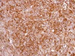
- Immunohistochemical analysis of paraffin-embedded CL1-0 xenograft, using GHS-R1 (Product # PA5-28752) antibody at 1:100 dilution. Antigen Retrieval: Citrate buffer, pH 6.0, 15 min.
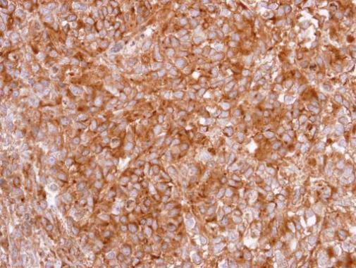
Supportive validation
- Submitted by
- Invitrogen Antibodies (provider)
- Main image

- Experimental details
- Fig. 1 Altered thymic architecture in mdx mice. Representative images of H&E staining of thymus of 3-month-old C57Bl and mdx mice revealed differences in medullary/cortex boundaries between animals (dashed white line) ( a ). WB analysis showed the downregulation of cytokeratin 14/16 in mdx thymus related to C57Bl ( b ). Thymic architecture of C57Bl and mdx mice characterized by immunofluorescence staining for cortical cytokeratin CK8 (green) and medullary cytokeratin CK5 (red) confirmed changes in dystrophic thymic environment. Graph displays fluorescence area % occupied by CK5 and CK8, as calculated by ImageJ software ( c ). Double immunofluorescence staining for CK5 (red) and CD3 (green) of C57Bl and mdx thymi portrayed a loosen embedding of CD3+ cells within dystrophic medulla. d Staining of ghrelin (GHR) and ghrelin receptor (GHS-R) (red) showed a comparable distribution of GHR between animals, but a prevalent expression of GHS-R in thymus of C57Bl mice. Of note, GHR was preferentially found in proximity of cortical CK8 (green). e Graph displays fluorescence area % occupied by GHS-R, as calculated by ImageJ software, in C57Bl and mdx mice ( f ). Expression of FoxP3+ cells (magenta) was evaluated by immunofluorescence staining within CK5+ thymic medulla (green). C-terminal containing dystrophin isoforms were detected by DYS-2 antibody (red) to identify a specific protein distribution within thymus. mTEC maturation level was evaluated by immunohistochemistry staining of C57