 Explore
 Validate
 Learn
Learn Western blot
 Immunocytochemistry
 Immunohistochemistry
Antibody data
- Antibody Data
- Antigen structure
- References [0]
- Comments [0]
- Validations
- Western blot [4]
- Immunocytochemistry [2]
Submit
Validation data
Reference
Comment
Report error
- Product number
- LS-C790719 - Provider product page

- Provider
- LSBio
- Product name
- ROS1 / ROS Antibody (aa2126-2347, clone OTI7E4, Carrier-free) LS-C790719
- Antibody type
- Monoclonal
- Description
- Purified from ascites.
- Reactivity
- Human
- Host
- Mouse
- Isotype
- IgG
- Antibody clone number
- OTI7E4
- Storage
- Store at -20°C. Avoid freeze-thaw cycles.
No comments: Submit comment
Enhanced validation
- Submitted by
- LSBio (provider)
- Enhanced method
- Genetic validation
- Main image

- Experimental details
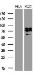
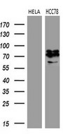
- Western blot analysis of extracts. (35ug) from 2 different cell lines by using anti-ROS1 monoclonal antibody. (1:500)

- Submitted by
- LSBio (provider)
- Enhanced method
- Genetic validation
- Main image
- Experimental details
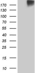
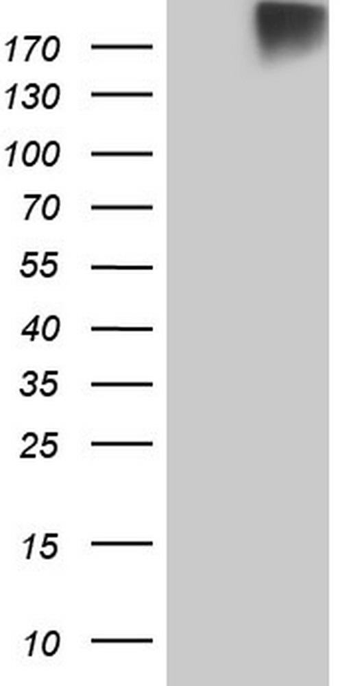
- HEK293T cells were transfected with the pCMV6-ENTRY control. (Left lane) or pCMV6-ENTRY ROS1. (Right lane) cDNA for 48 hrs and lysed. Equivalent amounts of cell lysates. (5 ug per lane) were separated by SDS-PAGE and immunoblotted with anti-ROS1.

- Submitted by
- LSBio (provider)
- Main image

- Experimental details
- HEK293T cells were transfected with the pCMV6-ENTRY control. (Left lane) or pCMV6-ENTRY ROS1. (Right lane) cDNA for 48 hrs and lysed. Equivalent amounts of cell lysates. (5 ug per lane) were separated by SDS-PAGE and immunoblotted with anti-ROS1.
- Submitted by
- LSBio (provider)
- Main image
- Experimental details
- Western blot analysis of extracts. (35ug) from 2 different cell lines by using anti-ROS1 monoclonal antibody. (1:500)
Supportive validation
- Submitted by
- LSBio (provider)
- Enhanced method
- Genetic validation
- Main image
- Experimental details
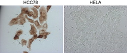
- Immunocytochemistry staining of HCC78 cells using anti-ROS1 mouse monoclonal antibody. (Left). The right is HELA cells as negative control. (1:2000)

- Submitted by
- LSBio (provider)
- Main image

- Experimental details
- Immunocytochemistry staining of HCC78 cells using anti-ROS1 mouse monoclonal antibody. (Left). The right is HELA cells as negative control. (1:2000)
