Explore
 Validate
 Learn
Learn Western blot
Antibody data
- Antibody Data
- Antigen structure
- References [0]
- Comments [0]
- Validations
- Western blot [2]
- Immunocytochemistry [1]
Submit
Validation data
Reference
Comment
Report error
- Product number
- LS-C776954 - Provider product page

- Provider
- LSBio
- Product name
- HYOU1 / ORP150 Antibody (clone 6E3-2C3) LS-C776954
- Antibody type
- Monoclonal
- Description
- Protein G purified
- Reactivity
- Human, Mouse, Rat
- Host
- Mouse
- Isotype
- IgG
- Antibody clone number
- 6E3-2C3
- Storage
- Store at -20°C.
No comments: Submit comment
Supportive validation
- Submitted by
- LSBio (provider)
- Main image

- Experimental details
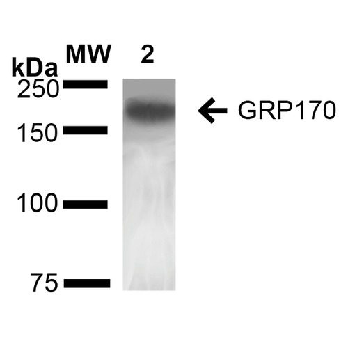
- Western Blot analysis of Rat Liver showing detection of ~170 kDa GRP170 protein using Mouse Anti-GRP170 Monoclonal Antibody, Clone 6E3-2C3. Lane 1: Molecular Weight Ladder (MW). Lane 2: Rat Liver cell lysate. Load: 20 µg. Block: 2% BSA and 2% Skim Milk in 1X TBST. Primary Antibody: Mouse Anti-GRP170 Monoclonal Antibody at 1:1000 for 16 hours at 4°C. Secondary Antibody: Goat Anti-Mouse IgG: HRP at 1:100 for 60 min at RT. Color Development: ECL solution for 6 min in RT. Predicted/Observed Size: ~170 kDa.
- Submitted by
- LSBio (provider)
- Main image
- Experimental details
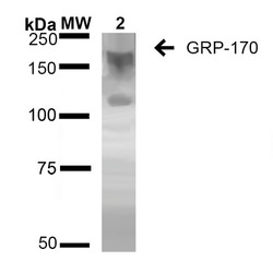
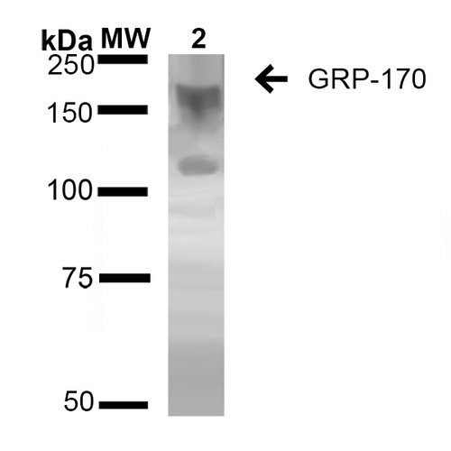
- Western Blot analysis of Human Embryonic kidney epithelial cell line (HEK293) lysates showing detection of ~170 kDa GRP170 protein using Mouse Anti-GRP170 Monoclonal Antibody, Clone 6E3-2C2. Lane 1: Molecular Weight Ladder (MW). Lane 2: HEK-293 cell lysate. Load: 20 µg. Block: 2% BSA and 2% Skim Milk in 1X TBST. Primary Antibody: Mouse Anti-GRP170 Monoclonal Antibody at 1:1000 for 16 hours at 4°C. Secondary Antibody: Goat Anti-Mouse IgG: HRP at 1:100 for 60 min at RT. Color Development: ECL solution for 6 min in RT. Predicted/Observed Size: ~170 kDa. Other Band(s): 100 kDa.
Supportive validation
- Submitted by
- LSBio (provider)
- Main image
- Experimental details
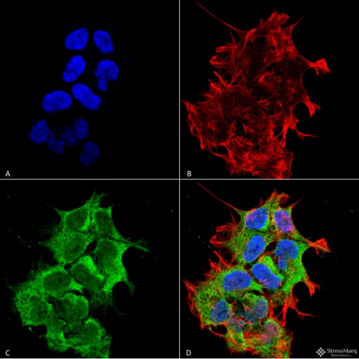
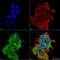
- Immunocytochemistry/Immunofluorescence analysis using Mouse Anti-GRP170 Monoclonal Antibody, Clone 6E3-2C3. Tissue: Neuroblastoma cell line (SK-N-BE). Species: Human. Fixation: 4% Formaldehyde for 15 min at RT. Primary Antibody: Mouse Anti-GRP170 Monoclonal Antibody at 1:100 for 60 min at RT. Secondary Antibody: Goat Anti-Mouse ATTO 488 at 1:100 for 60 min at RT. Counterstain: Phalloidin Texas Red F-Actin stain; DAPI (blue) nuclear stain at 1:1000; 1:5000 for 60 min RT, 5 min RT. Localization: Endoplasmic Reticulum, Endoplasmic Reticulum Lumen. Magnification: 60X. (A) DAPI (blue) nuclear stain. (B) Phalloidin Texas Red F-Actin stain. (C) GRP170 Antibody. (D) Composite.