Explore
 Validate
 Learn
Learn Western blot
 Immunocytochemistry
Antibody data
- Antibody Data
- Antigen structure
- References [1]
- Comments [0]
- Validations
- Immunocytochemistry [1]
- Immunohistochemistry [1]
Submit
Validation data
Reference
Comment
Report error
- Product number
- HPA019828 - Provider product page

- Provider
- Atlas Antibodies
- Proper citation
- Atlas Antibodies Cat#HPA019828, RRID:AB_1844801
- Product name
- Anti-AMPH
- Antibody type
- Polyclonal
- Description
- Polyclonal Antibody against Human AMPH, Gene description: amphiphysin, Validated applications: ICC, IHC, WB, Uniprot ID: P49418, Storage: Store at +4°C for short term storage. Long time storage is recommended at -20°C.
- Reactivity
- Human
- Host
- Rabbit
- Conjugate
- Unconjugated
- Isotype
- IgG
- Vial size
- 100 µl
- Concentration
- 0.1 mg/ml
- Storage
- Store at +4°C for short term storage. Long time storage is recommended at -20°C.
- Handling
- The antibody solution should be gently mixed before use.
Submitted references Association of CSF proteins with tau and amyloid β levels in asymptomatic 70-year-olds
Remnestål J, Bergström S, Olofsson J, Sjöstedt E, Uhlén M, Blennow K, Zetterberg H, Zettergren A, Kern S, Skoog I, Nilsson P, Månberg A
Alzheimer's Research & Therapy 2021;13(1)
Alzheimer's Research & Therapy 2021;13(1)
No comments: Submit comment
Supportive validation
- Submitted by
- Atlas Antibodies (provider)
- Main image

- Experimental details
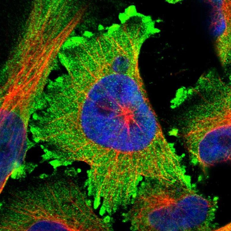
- Immunofluorescent staining of human cell line U-251 MG shows localization to plasma membrane & cytosol.
- Sample type
- Human
Supportive validation
- Submitted by
- Atlas Antibodies (provider)
- Enhanced method
- Orthogonal validation
- Main image
- Experimental details
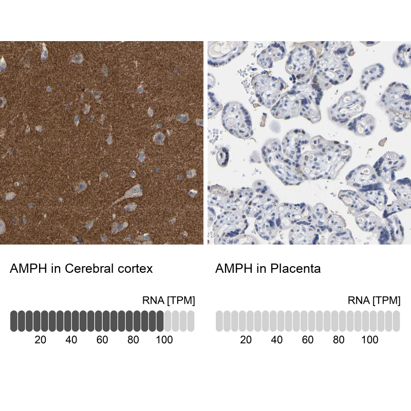
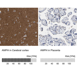
- Immunohistochemistry analysis in human cerebral cortex and placenta tissues using HPA019828 antibody. Corresponding AMPH RNA-seq data are presented for the same tissues.
- Sample type
- Human
- Protocol
- Protocol