Explore
 Validate
 Learn
Learn Western blot
 ELISA
ELISAAntibody data
- Antibody Data
- Antigen structure
- References [0]
- Comments [0]
- Validations
- Western blot [4]
- Immunohistochemistry [11]
Submit
Validation data
Reference
Comment
Report error
- Product number
- ABIN2506520 - Provider product page

- Provider
- antibodies-online
- Product name
- anti-Testis-Specific Kinase 2 (TESK2) antibody
- Antibody type
- Polyclonal
- Antigen
- The antiserum was produced against synthesized peptide derived from internal of human TESK2.
- Description
- The antibody was affinity-purified from rabbit antiserum by affinity-chromatography using epitope-specific immunogen.
- Reactivity
- Human, Mouse, Rat
- Host
- Rabbit
- Isotype
- IgG
- Vial size
- 100 μg
- Storage
- Store at -20°C/1 year
No comments: Submit comment
Supportive validation
- Submitted by
- antibodies-online (provider)
- Main image
- Experimental details
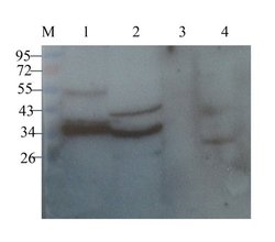
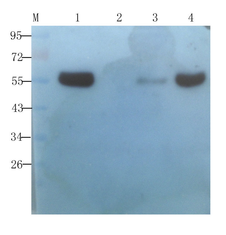
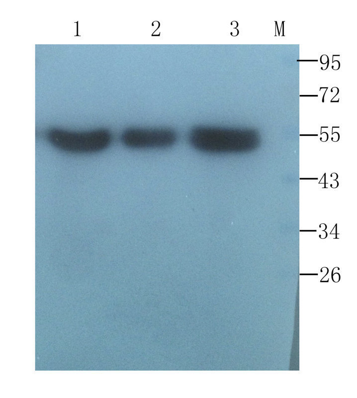
- Western blot analysis of rat thymus (lane 1), rat liver (lane 2), mouse spleen (lane 3), mouse samll intestine (lane 4) using CD4 antibody (1 ug/ml)

- Submitted by
- antibodies-online (provider)
- Main image

- Experimental details
- WB analysis of human breast cancer (lane 1), human thyroid cancer (lane 2), human endometrial cancer (lane 3), human ovarian cancer (lane 4) using CD4 antibody (1 ug/ml)
- Submitted by
- antibodies-online (provider)
- Main image

- Experimental details
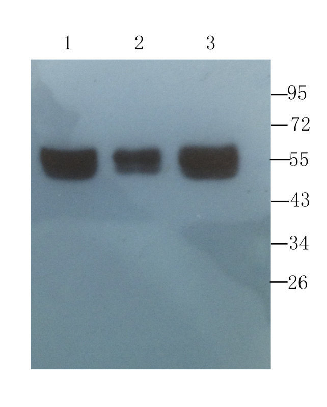
- Western blot analysis of human breast tumor (lane 1), human mammary fibroma (lane 2), human breast cancer (lane 3) using CD4 antibody (1 ug/ml)
- Submitted by
- antibodies-online (provider)
- Main image

- Experimental details
- WB analysis of human breast tumor (lane 1), human mammary fibroma (lane 2), human breast cancer (lane 3) using CD4 antibody (1 ug/ml)
Supportive validation
- Submitted by
- antibodies-online (provider)
- Main image

- Experimental details
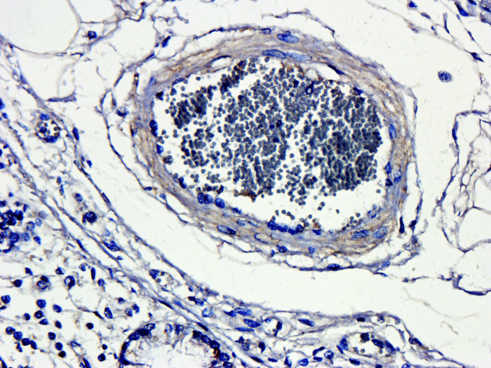
- Immunohistochemical staining of pig large intestines tissue using anti-CD4 (dilution of primary antibody - 1:200)

- Submitted by
- antibodies-online (provider)
- Main image

- Experimental details
- IHC-P staining of rat colon tissue using CD4 antibody (5 ug/ml)

- Submitted by
- antibodies-online (provider)
- Main image

- Experimental details
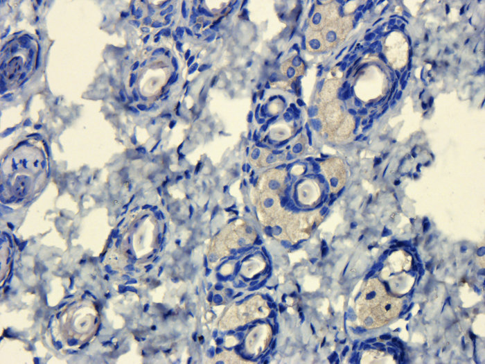
- Immunohistochemical staining of rat skin tissue using anti-CD4 (2.5 ug/ml)

- Submitted by
- antibodies-online (provider)
- Main image

- Experimental details
- Immunohistochemical staining of mouse lymph node tissue using anti-CD4 (5 ug/ml)
- Submitted by
- antibodies-online (provider)
- Main image
- Experimental details
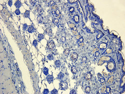
- Immunohistochemical staining of paraffin embedded mouse skin tissue using CD4 antibody (2.5 ug/ml)
- Submitted by
- antibodies-online (provider)
- Main image
- Experimental details
- Immunohistochemical staining of paraffin embedded mouse skin tissue using CD4 antibody (2.5 ug/ml)
- Submitted by
- antibodies-online (provider)
- Main image
- Experimental details
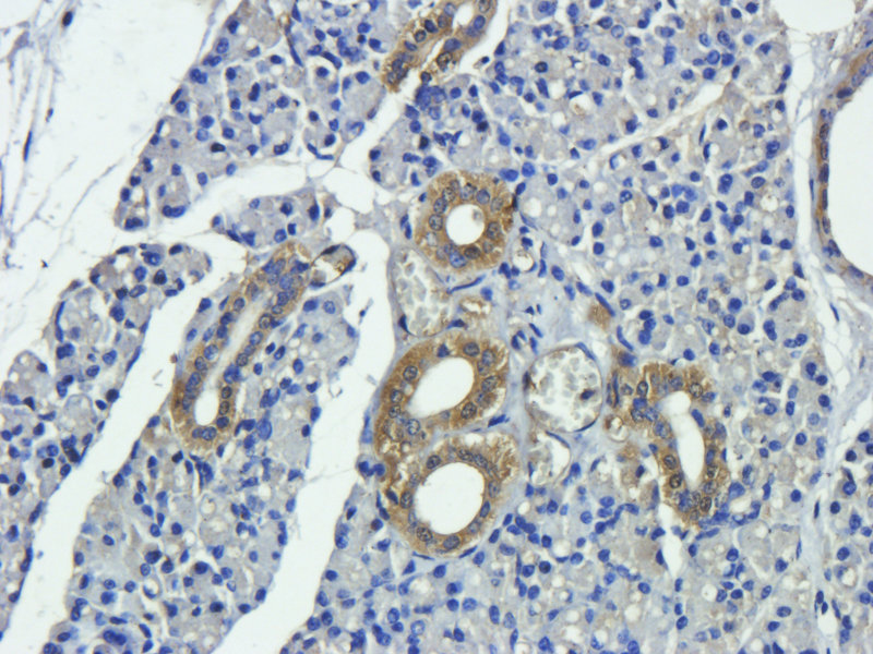
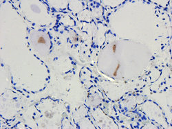
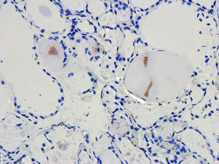
- IHC-P image of human thyroid carcinoma tissue using CD4 antibody (2.5 ug/ml)

- Submitted by
- antibodies-online (provider)
- Main image
- Experimental details
- IHC-P staining of human thyroid carcinoma tissue using CD4 antibody (2.5 ug/ml)
- Submitted by
- antibodies-online (provider)
- Main image
- Experimental details
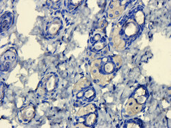
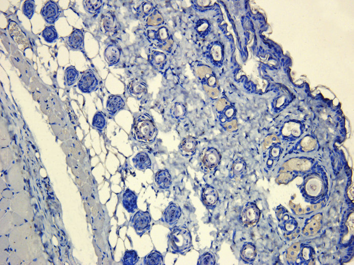
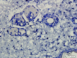
- IHC-P image of rat skin tissue using CD4 antibody (2.5 ug/ml)

- Submitted by
- antibodies-online (provider)
- Main image
- Experimental details
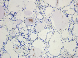
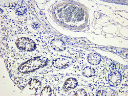
- Immunohistochemical staining of paraffin embedded pig large intestines tissue using CD4 antibody (2.5 ug/ml)

- Submitted by
- antibodies-online (provider)
- Main image

- Experimental details
- IHC-P image of pig large intestines tissue using anti-CD4 (2.5 ug/ml)