 Explore
 Validate
 Learn
Learn Western blot
 ELISA
ELISAAntibody data
- Antibody Data
- Antigen structure
- References [0]
- Comments [0]
- Validations
- Western blot [2]
- Immunocytochemistry [3]
- Immunohistochemistry [9]
Submit
Validation data
Reference
Comment
Report error
- Product number
- RQ7222 - Provider product page

- Provider
- NSJ Bioreagents
- Product name
- COX5B Antibody / Cytochrome c oxidase subunit 5B
- Antibody type
- Polyclonal
- Description
- This highly specific COX5B antibody is suitable for use in Western blot/Immunohistochemistry/Immunofluorescence/Flow cytometry/Direct ELISA applications with human, mouse and rat samples.
- Reactivity
- Human, Mouse, Rat
- Host
- Rabbit
- Conjugate
- Unconjugated
- Vial size
- 100 ug
- Concentration
- 0.5mg/ml if reconstituted with 0.2ml sterile DI water
- Storage
- After reconstitution, the COX5B antibody can be stored for up to one month at 4oC. For long-term, aliquot and store at -20oC. Avoid repeated freezing and thawing.
No comments: Submit comment
Supportive validation
- Submitted by
- NSJ Bioreagents (provider)
- Main image
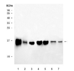
- Experimental details
- Western blot testing of 1) rat heart, 2) rat liver, 3) rat brain, 4) mouse heart, 5) mouse liver, 6) mouse brain and 7) mouse lung tissue lysate with COX5B antibody. Predicted molecular weight ~14 kDa.

- Submitted by
- NSJ Bioreagents (provider)
- Main image

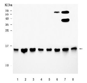
- Experimental details
- Western blot testing of human 1) HeLa, 2) HepG2, 3) PC-3, 4) 293T, 5) Caco-2, 6) MCF7, 7) HCCT and 8) HCCP cell lysate with COX5B antibody. Predicted molecular weight ~14 kDa.
Supportive validation
- Submitted by
- NSJ Bioreagents (provider)
- Main image
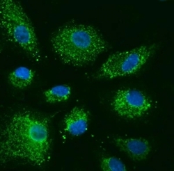
- Experimental details
- Immunofluorescent staining of FFPE human A549 cells with COX5B antibody (green) and DAPI nuclear stain (blue). HIER: steam section in pH6 citrate buffer for 20 min.
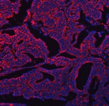
- Submitted by
- NSJ Bioreagents (provider)
- Main image
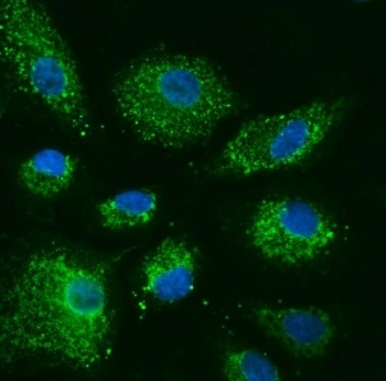
- Experimental details
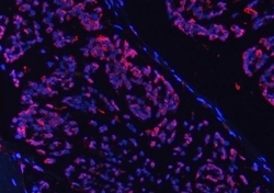
- Immunofluorescent staining of FFPE human breast cancer tissue with COX5B antibody (red) and DAPI nuclear stain (blue). HIER: steam section in pH8 EDTA buffer for 20 min.

- Submitted by
- NSJ Bioreagents (provider)
- Main image

- Experimental details
- Immunofluorescent staining of FFPE human ovarian cancer tissue with COX5B antibody (red) and DAPI nuclear stain (blue). HIER: steam section in pH8 EDTA buffer for 20 min.
Supportive validation
- Submitted by
- NSJ Bioreagents (provider)
- Main image
- Experimental details
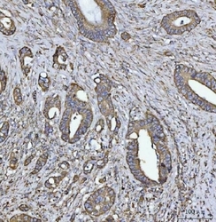
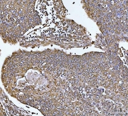
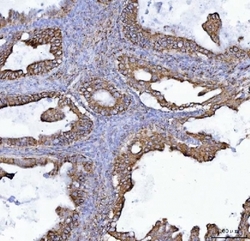
- IHC staining of FFPE human breast cancer tissue with COX5B antibody. HIER: boil tissue sections in pH8 EDTA for 20 min and allow to cool before testing.
- Submitted by
- NSJ Bioreagents (provider)
- Main image
- Experimental details
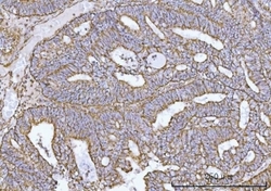
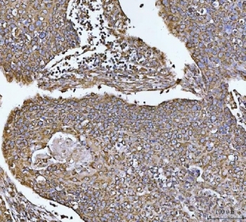
- IHC staining of FFPE human colorectal adenocarcinoma tissue with COX5B antibody. HIER: boil tissue sections in pH8 EDTA for 20 min and allow to cool before testing.
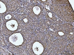
- Submitted by
- NSJ Bioreagents (provider)
- Main image
- Experimental details
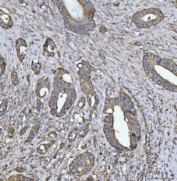
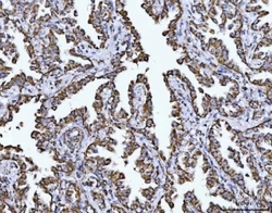
- IHC staining of FFPE human endometrial carcinoma tissue with COX5B antibody. HIER: boil tissue sections in pH8 EDTA for 20 min and allow to cool before testing.
- Submitted by
- NSJ Bioreagents (provider)
- Main image
- Experimental details
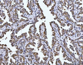
- IHC staining of FFPE human esophageal squamous carcinoma tissue with COX5B antibody. HIER: boil tissue sections in pH8 EDTA for 20 min and allow to cool before testing.
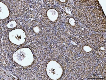
- Submitted by
- NSJ Bioreagents (provider)
- Main image
- Experimental details
- IHC staining of FFPE human lung cancer tissue with COX5B antibody. HIER: boil tissue sections in pH8 EDTA for 20 min and allow to cool before testing.
- Submitted by
- NSJ Bioreagents (provider)
- Main image
- Experimental details
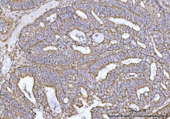
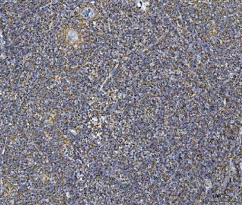
- IHC staining of FFPE human ovarian cancer tissue with COX5B antibody. HIER: boil tissue sections in pH8 EDTA for 20 min and allow to cool before testing.

- Submitted by
- NSJ Bioreagents (provider)
- Main image
- Experimental details
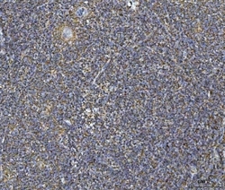
- IHC staining of FFPE human tonsil tissue with COX5B antibody. HIER: boil tissue sections in pH8 EDTA for 20 min and allow to cool before testing.
- Submitted by
- NSJ Bioreagents (provider)
- Main image
- Experimental details
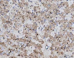
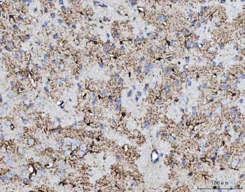
- IHC staining of FFPE mouse brain tissue with COX5B antibody. HIER: boil tissue sections in pH8 EDTA for 20 min and allow to cool before testing.
- Submitted by
- NSJ Bioreagents (provider)
- Main image
- Experimental details
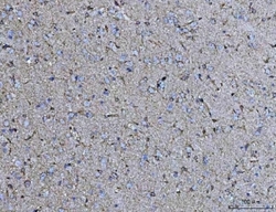
- IHC staining of FFPE rat brain tissue with COX5B antibody. HIER: boil tissue sections in pH8 EDTA for 20 min and allow to cool before testing.
