 Explore
 Validate
 Learn
Learn Western blot
Antibody data
- Antibody Data
- Antigen structure
- References [2]
- Comments [0]
- Validations
- Western blot [1]
- Immunocytochemistry [2]
- Immunohistochemistry [4]
Submit
Validation data
Reference
Comment
Report error
- Product number
- GTX132974 - Provider product page

- Provider
- GeneTex
- Product name
- NeuN antibody
- Antibody type
- Polyclonal
- Reactivity
- Mouse, Rat
- Host
- Rabbit
Submitted references Anti-TNF-α restricts dengue virus-induced neuropathy.
Radioprotective effect of ursolic acid in radiation-induced impairment of neurogenesis, learning and memory in adolescent BALB/c mouse.
Jhan MK, HuangFu WC, Chen YF, Kao JC, Tsai TT, Ho MR, Shen TJ, Tseng PC, Wang YT, Lin CF
Journal of leukocyte biology 2018 Nov;104(5):961-968
Journal of leukocyte biology 2018 Nov;104(5):961-968
Radioprotective effect of ursolic acid in radiation-induced impairment of neurogenesis, learning and memory in adolescent BALB/c mouse.
Tang FR, Loke WK, Wong P, Khoo BC
Physiology & behavior 2017 Jun 1;175:37-46
Physiology & behavior 2017 Jun 1;175:37-46
No comments: Submit comment
Supportive validation
- Submitted by
- GeneTex (provider)
- Main image
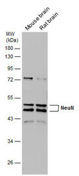
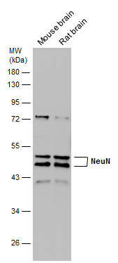
- Experimental details
- Various tissue extracts (50 ?g) were separated by 10% SDS-PAGE, and the membrane was blotted with NeuN antibody (GTX132974) diluted at 1:1000.
Supportive validation
- Submitted by
- GeneTex (provider)
- Main image
- Experimental details
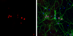
- NeuN antibody detects NeuN protein at nucleus by immunofluorescent analysis.Sample: Rat E18 primary cortical neuron, DIV 8 cells were fixed in 4% paraformaldehyde at RT for 15 min.Red: NeuN protein stained by NeuN antibody (GTX132974) diluted at 1:250.Green: alpha Tubulin, stained by alpha Tubulin antibody [GT114] (GTX628802) diluted at 1:250.Blue: Fluoroshield with DAPI (GTX30920).

- Submitted by
- GeneTex (provider)
- Main image
- Experimental details
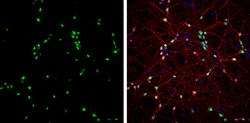
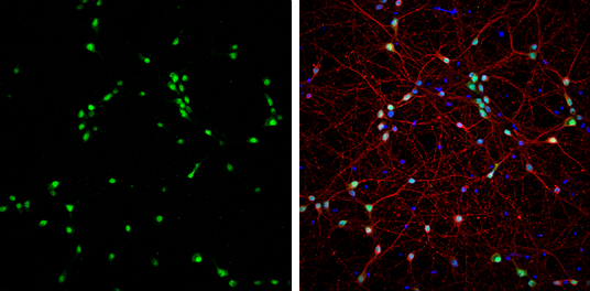
- NeuN antibody detects NeuN protein at nucleus by immunofluorescent analysis.Sample: DIV9 rat E18 primary cortical neurons were fixed in 4% paraformaldehyde at RT for 15 min.Green: NeuN protein stained by NeuN antibody (GTX132974) diluted at 1:1000.Red: beta Tubulin 3/ Tuj1, stained by beta Tubulin 3/ Tuj1 antibody [GT886] (GTX631830) diluted at 1:500.Blue: Fluoroshield with DAPI (GTX30920).
Supportive validation
- Submitted by
- GeneTex (provider)
- Main image
- Experimental details
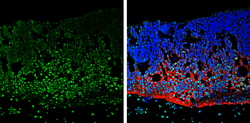
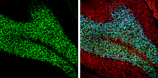
- NeuN antibody detects NeuN protein at nucleus by immunohistochemical analysis.Sample: Frozen sectioned E13.5 rat brain. Green: NeuN protein stained by NeuN antibody (GTX132974) diluted at 1:250.Red: beta Tubulin 3/ TUJ1, a mature neuron marker, stained by beta Tubulin 3/ TUJ1 antibody [GT11710] (GTX631836) diluted at 1:250.Blue: Fluoroshield with DAPI (GTX30920).
- Submitted by
- GeneTex (provider)
- Main image

- Experimental details
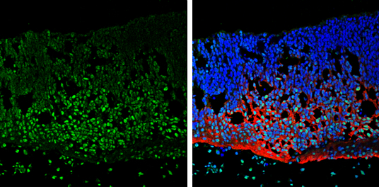
- NeuN antibody detects NeuN protein by immunohistochemical analysis.Sample: Frozen sectioned adult mouse retina. Green: NeuN protein stained by NeuN antibody (GTX132974) diluted at 1:250.Red: Protein kinase C alpha staining.Blue: Fluoroshield with DAPI (GTX30920).
- Submitted by
- GeneTex (provider)
- Main image

- Experimental details
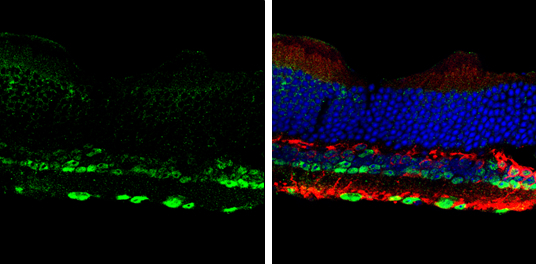
- NeuN antibody detects NeuN protein expression by immunohistochemical analysis.Sample: Frozen-sectioned adult mouse cerebellum. Green: NeuN protein stained by NeuN antibody (GTX132974) diluted at 1:250.Red: beta Tubulin 3/ TUJ1, stained by beta Tubulin 3/ TUJ1 antibody [GT11710] (GTX631836) diluted at 1:500.Blue: Fluoroshield with DAPI (GTX30920).
- Submitted by
- GeneTex (provider)
- Main image
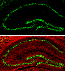
- Experimental details
- NeuN antibody detects NeuN protein expression by immunohistochemical analysis.Sample: Frozen-sectioned adult mouse hippocampus. Green: NeuN protein stained by NeuN antibody (GTX132974) diluted at 1:250.Red: alpha Tubulin, stained by alpha Tubulin antibody [GT114] (GTX628802) diluted at 1:500.
