Explore
 Validate
 Learn
Learn Western blot
Antibody data
- Antibody Data
- Antigen structure
- References [8]
- Comments [0]
- Validations
- Western blot [4]
- Immunocytochemistry [3]
- Immunoprecipitation [1]
- Immunohistochemistry [2]
- Flow cytometry [1]
- Other assay [3]
Submit
Validation data
Reference
Comment
Report error
- Product number
- 702022 - Provider product page

- Provider
- Invitrogen Antibodies
- Product name
- NeuN Recombinant Rabbit Monoclonal Antibody (14H6L24)
- Antibody type
- Monoclonal
- Antigen
- Other
- Description
- This antibody is predicted to react with Monkey, Rat and Mouse. Recombinant rabbit monoclonal antibodies are produced using in vitro expression systems. The expression systems are developed by cloning in the specific antibody DNA sequences from immunoreactive rabbits. Then, individual clones are screened to select the best candidates for production. The advantages of using recombinant rabbit monoclonal antibodies include: better specificity and sensitivity, lot-to-lot consistency, animal origin-free formulations, and broader immunoreactivity to diverse targets due to larger rabbit immune repertoire.
- Reactivity
- Human, Mouse, Rat
- Host
- Rabbit
- Isotype
- IgG
- Antibody clone number
- 14H6L24
- Vial size
- 100 μg
- Concentration
- 0.5 mg/mL
- Storage
- Store at 4°C short term. For long term storage, store at -20°C, avoiding freeze/thaw cycles.
Submitted references Molecular Markers of Mechanosensation in Glycinergic Neurons in the Avian Lumbosacral Spinal Cord.
Aberrant crosstalk between insulin signaling and mTOR in young Down syndrome individuals revealed by neuronal-derived extracellular vesicles.
Small molecules efficiently reprogram apical papilla stem cells into neuron-like cells.
Developmental Regulation of Homeostatic Plasticity in Mouse Primary Visual Cortex.
Ultrashort Wave Combined with Human Umbilical Cord Mesenchymal Stem Cell (HUC-MSC) Transplantation Inhibits NLRP3 Inflammasome and Improves Spinal Cord Injury via MK2/TTP Signalling Pathway.
Fgr contributes to hemorrhage-induced thalamic pain by activating NF-κB/ERK1/2 pathways.
Inflammasome Activation Induces Pyroptosis in the Retina Exposed to Ocular Hypertension Injury.
Gemfibrozil, a Lipid-Lowering Drug, Lowers Amyloid Plaque Pathology and Enhances Memory in a Mouse Model of Alzheimer's Disease via Peroxisome Proliferator-Activated Receptor α.
Stanchak KE, Miller KE, Lumsden EW, Shikiar D, Davis C, Brunton BW, Perkel DJ
eNeuro 2022 Sep-Oct;9(5)
eNeuro 2022 Sep-Oct;9(5)
Aberrant crosstalk between insulin signaling and mTOR in young Down syndrome individuals revealed by neuronal-derived extracellular vesicles.
Perluigi M, Picca A, Montanari E, Calvani R, Marini F, Matassa R, Tramutola A, Villani A, Familiari G, Domenico FD, Butterfield DA, Oh KJ, Marzetti E, Valentini D, Barone E
Alzheimer's & dementia : the journal of the Alzheimer's Association 2022 Aug;18(8):1498-1510
Alzheimer's & dementia : the journal of the Alzheimer's Association 2022 Aug;18(8):1498-1510
Small molecules efficiently reprogram apical papilla stem cells into neuron-like cells.
Chen Q, Yuan C, Jiang S, Heng BC, Zou T, Shen Z, Wang P, Zhang C
Experimental and therapeutic medicine 2021 Jun;21(6):546
Experimental and therapeutic medicine 2021 Jun;21(6):546
Developmental Regulation of Homeostatic Plasticity in Mouse Primary Visual Cortex.
Wen W, Turrigiano GG
The Journal of neuroscience : the official journal of the Society for Neuroscience 2021 Dec 1;41(48):9891-9905
The Journal of neuroscience : the official journal of the Society for Neuroscience 2021 Dec 1;41(48):9891-9905
Ultrashort Wave Combined with Human Umbilical Cord Mesenchymal Stem Cell (HUC-MSC) Transplantation Inhibits NLRP3 Inflammasome and Improves Spinal Cord Injury via MK2/TTP Signalling Pathway.
Na L, Wang S, Liu T, Zhang L
BioMed research international 2020;2020:3021750
BioMed research international 2020;2020:3021750
Fgr contributes to hemorrhage-induced thalamic pain by activating NF-κB/ERK1/2 pathways.
Huang T, Fu G, Gao J, Zhang Y, Cai W, Wu S, Jia S, Xia S, Bachmann T, Bekker A, Tao YX
JCI insight 2020 Oct 15;5(20)
JCI insight 2020 Oct 15;5(20)
Inflammasome Activation Induces Pyroptosis in the Retina Exposed to Ocular Hypertension Injury.
Pronin A, Pham D, An W, Dvoriantchikova G, Reshetnikova G, Qiao J, Kozhekbaeva Z, Reiser AE, Slepak VZ, Shestopalov VI
Frontiers in molecular neuroscience 2019;12:36
Frontiers in molecular neuroscience 2019;12:36
Gemfibrozil, a Lipid-Lowering Drug, Lowers Amyloid Plaque Pathology and Enhances Memory in a Mouse Model of Alzheimer's Disease via Peroxisome Proliferator-Activated Receptor α.
Chandra S, Pahan K
Journal of Alzheimer's disease reports 2019 May 18;3(1):149-168
Journal of Alzheimer's disease reports 2019 May 18;3(1):149-168
No comments: Submit comment
Supportive validation
- Submitted by
- Invitrogen Antibodies (provider)
- Main image

- Experimental details
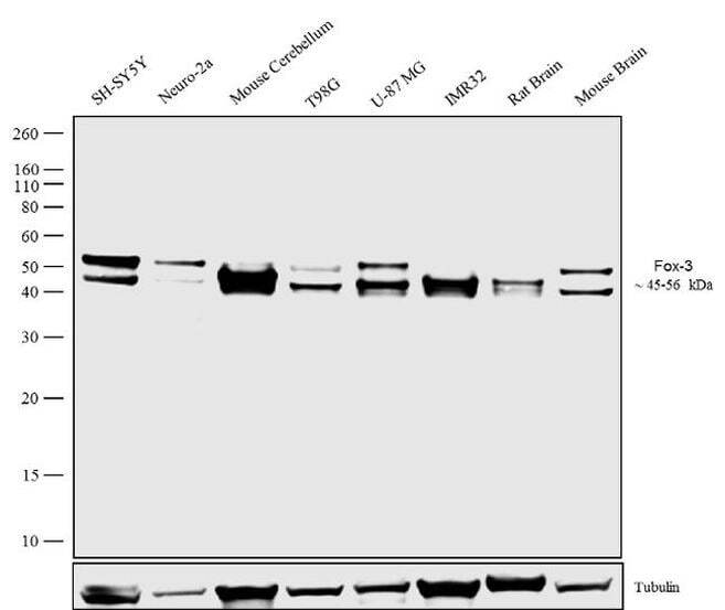
- Western blot analysis was performed on whole cell extracts (30 µg lysate) SH-SY5Y (Lane 1), Neuro-2a (Lane 2), Mouse cerebellum (Lane 3), T98G (Lane 4), U-87 MG (Lane 5), IMR32 (Lane 6), Rat Brain (Lane 7), Mouse Brain (Lane 8). The blots were probed with Anti- Fox3 Recombinant Rabbit Monoclonal Antibody (Product # 702022, 1-2 µg/mL) and detected by chemiluminescence using Goat anti-Rabbit IgG (Heavy Chain) Superclonal™ Secondary Antibody, HRP conjugate (Product # A27036, 0.4 µg/mL, 1:2500 dilution). A ~45 and 56 kDa band corresponding to Fox3 protein was observed in all cell lines. Known quantity of protein samples were electrophoresed using Novex® NuPAGE® 10% Bis-Tris gel (Product # NP0301BOX), XCell SureLock™ Electrophoresis System (Product # EI0002) and Novex® Sharp Pre-Stained Protein Standard (Product # LC5800). Resolved proteins were then transferred onto a nitrocellulose membrane with overnight wet transfer system. The membrane was probed with the relevant primary and secondary Antibody following blocking with 5% skimmed milk. Chemiluminescent detection was performed using Pierce™ ECL Western blotting Substrate (Product # 32106).
- Submitted by
- Invitrogen Antibodies (provider)
- Main image
- Experimental details
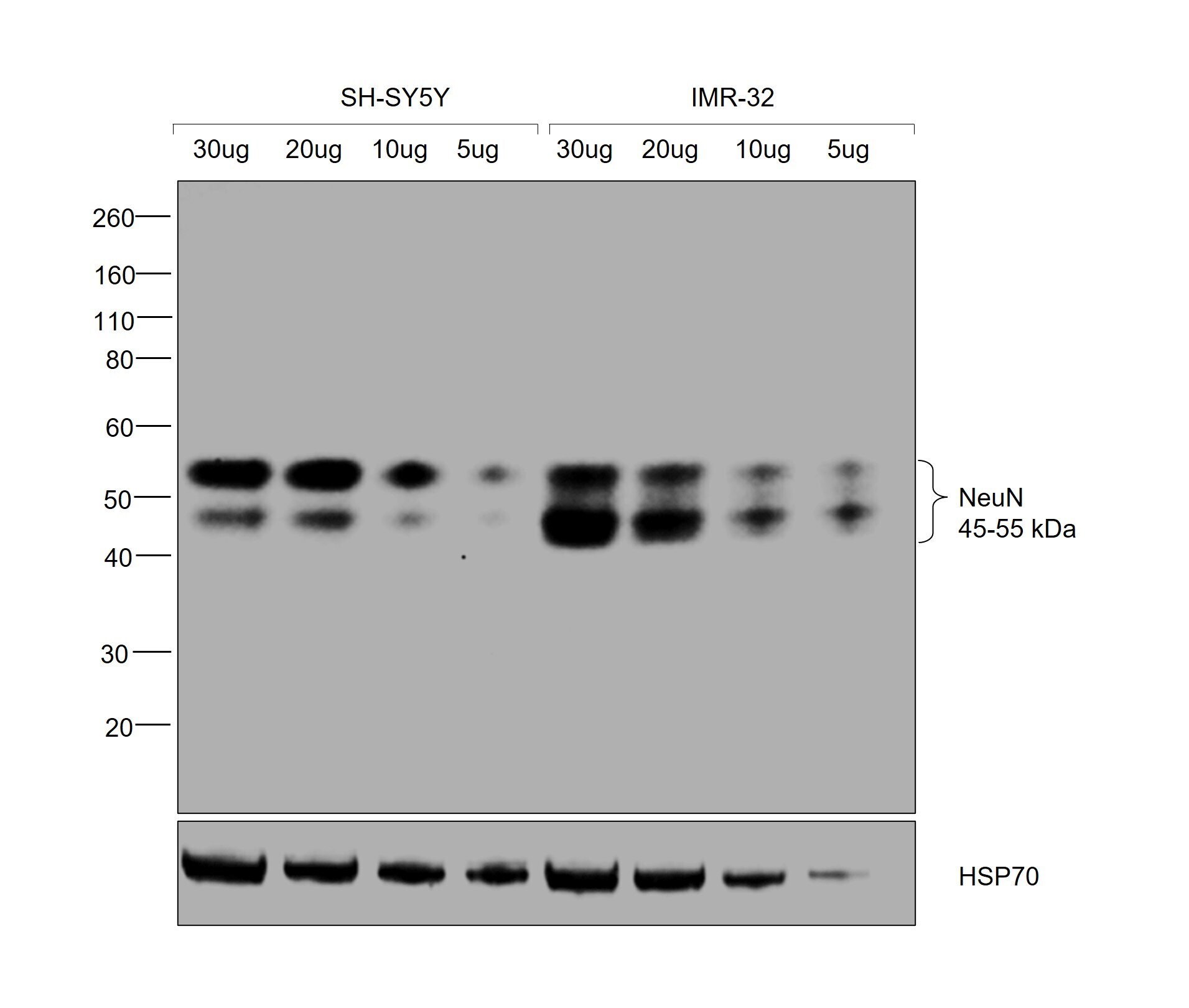
- Western blot was performed using NeuN Recombinant Rabbit Monoclonal Antibody (14H6L24) (Product # 702022) and bands between 45 and 55 kDa corresponding to NeuN were observed in varied concentrations of SH-SY5Y and IMR-32. Modified whole cell extracts (1% SDS) of SH-SY5Y (Lanes 1-4) and IMR-32 (Lane 5-8) loaded at different concentrations 30 µg, 20 µg, 10 µg and 5 µg were electrophoresed using NuPAGE™ 10% Bis-Tris gel (Product # NP0301BOX), 10 well. Resolved proteins were then transferred onto a nitrocellulose membrane (Product # IB33001) by iBlot™ 3 Western Blot Transfer Device (Product # IB31001). The blot was probed with the primary antibody (0.5 µg/mL) and detected by chemiluminescence with Goat anti-Rabbit IgG (H+L) Superclonal™ Recombinant Secondary Antibody, HRP (Product # A27036, 1:20,000) using the iBright™ FL1500 Imaging System (Product # A44115). Chemiluminescent detection was performed using SuperSignal™ West Pico PLUS Chemiluminescent Substrate (Product # 34580).
- Submitted by
- Invitrogen Antibodies (provider)
- Main image
- Experimental details
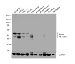
- Western blot was performed using NeuN Recombinant Rabbit Monoclonal Antibody (14H6L24) (Product # 702022) and bands between 45 and 55 kDa corresponding to NeuN were observed in all tested cell lines and tissues, except Mouse heart, Mouse kidney, Rat heart and Rat kidney which are known to be low expressors. Modified whole cell extracts (1% SDS) (30 µg lysate) of SH-SY5Y (Lane 1), IMR-32 (Lane 2), C8-D1A (Lane 3), RSC96 (Lane 4) and tissue lysates (30 µg lysate) of Mouse Brain (Lane 5), Mouse Heart (Lane 6), Mouse Kidney (Lane 7), Rat Brain (Lane 8), Rat Heart (Lane 9) and Rat Kidney (Lane 10) were electrophoresed using NuPAGE™ 10% Bis-Tris gel (Product # NP0301BOX), 12 well. Resolved proteins were then transferred onto a nitrocellulose membrane (Product # IB33001) by iBlot™ 3 Western Blot Transfer Device (Product # IB31001). The blot was probed with the primary antibody (1 µg/mL) and detected by chemiluminescence with Goat anti-Rabbit IgG (H+L) Superclonal™ Recombinant Secondary Antibody, HRP (Product # A27036, 1:20,000) using the iBright™ FL1500 Imaging System (Product # A44115). Chemiluminescent detection was performed using SuperSignal™ West Pico PLUS Chemiluminescent Substrate (Product # 34580).
- Submitted by
- Invitrogen Antibodies (provider)
- Main image
- Experimental details
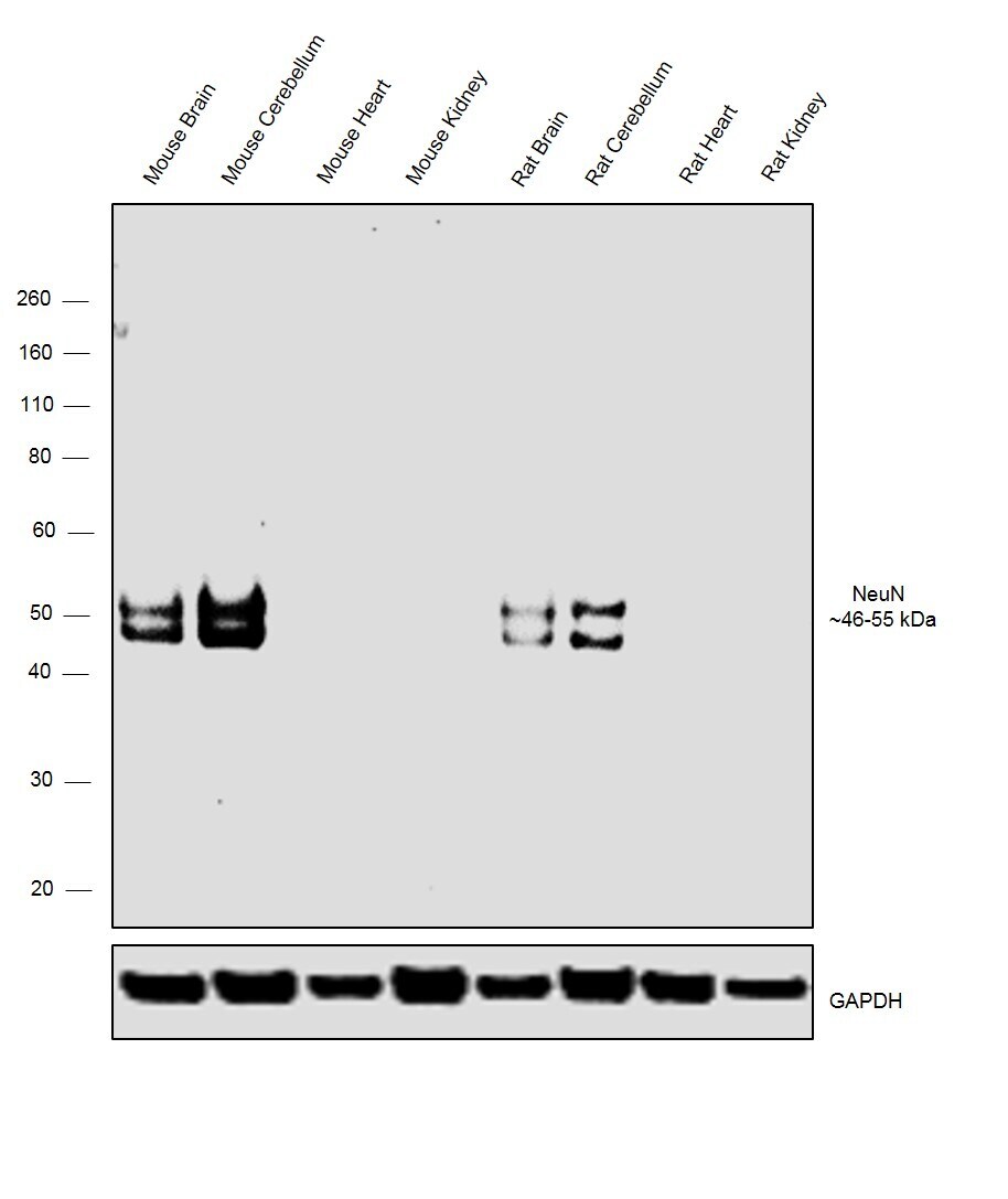
- Western blot was performed using Anti-NeuN Monoclonal Antibody, (Product # 702022) and bands at ~46-55 kDa corresponding to NeuN were observed across the Mouse and Rat tissues tested except Heart and Kidney which are reported to be negative. Tissue extracts (30 µg lysate) of Mouse Brain (Lane 1), Mouse Cerebellum (Lane 2), Mouse Heart (Lane 3), Mouse Kidney (Lane 4), Rat Brain (Lane 5), Rat Cerebellum (Lane 6), Rat Heart (Lane 7) and Rat Kidney (Lane 8) were electrophoresed using Novex® NuPAGE® 4-12% % Bis-Tris gel (Product # NP0322BOX). Resolved proteins were then transferred onto a nitrocellulose membrane (Product # IB23001) by iBlot® 2 Dry Blotting System (Product # IB21001). The blot was probed with the primary antibody (2 µg/mL) and detected by chemiluminescence with Goat anti- Rabbit IgG (Heavy Chain) Superclonal™ Recombinant Secondary Antibody, HRP (Product # A27036, 1:4,000 dilution) using the iBright FL 1000 (Product # A32752). Chemiluminescent detection was performed using Novex® ECL Chemiluminescent Substrate Reagent Kit (Product # WP20005).
Supportive validation
- Submitted by
- Invitrogen Antibodies (provider)
- Main image
- Experimental details
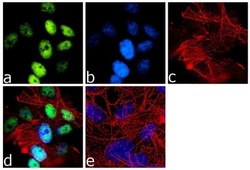
- Immunofluorescence was performed on fixed and permeabilized SH-SY5Y cells for detection of endogenous Fox3 using Anti- Fox3 Recombinant Rabbit Monoclonal Antibody (Product # 702022, 2 µg/mL) and labeled with Goat anti-Rabbit IgG (H+L) Superclonal™ Secondary Antibody, Alexa Fluor® 488 conjugate (Product # A27034, 1:2000). Panel a) shows representative cells that were stained for detection and localization of Fox3 protein (green), Panel b) is stained for nuclei (blue) using SlowFade® Gold Antifade Mountant with DAPI (Product # S36938). Panel c) represents cytoskeletal F-actin staining using Alexa Fluor® 555 Rhodamine Phalloidin (Product # R415, 1:300). Panel d) is a composite image of Panels a, b and c clearly demonstrating nuclear localization of Fox3. Panel e) represents control cells with no primary antibody to assess background.

- Submitted by
- Invitrogen Antibodies (provider)
- Main image
- Experimental details
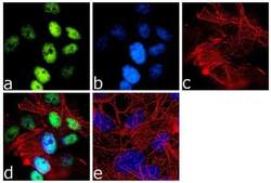
- Immunofluorescence was performed on fixed and permeabilized SH-SY5Y cells for detection of endogenous Fox3 using Anti- Fox3 Recombinant Rabbit Monoclonal Antibody (Product # 702022, 2 µg/mL) and labeled with Goat anti-Rabbit IgG (Heavy Chain) Superclonal™ Secondary Antibody, Alexa Fluor® 488 conjugate (Product # A27034, 1:2000). Panel a) shows representative cells that were stained for detection and localization of Fox3 protein (green), Panel b) is stained for nuclei (blue) using SlowFade® Gold Antifade Mountant with DAPI (Product # S36938). Panel c) represents cytoskeletal F-actin staining using Alexa Fluor® 555 Rhodamine Phalloidin (Product # R415, 1:300). Panel d) is a composite image of Panels a, b and c clearly demonstrating nuclear localization of Fox3. Panel e) represents control cells with no primary antibody to assess background.

- Submitted by
- Invitrogen Antibodies (provider)
- Main image
- Experimental details
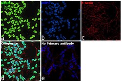
- Immunofluorescence analysis of NeuN was performed using 70% confluent log phase SH-SY5Y cells. The cells were fixed with 4% paraformaldehyde for 10 minutes, permeabilized with 0.1% Triton™ X-100 for 15 minutes and blocked with 2% BSA for 45 minutes at room temperature. The cells were labelled with NeuN Recombinant Rabbit Monoclonal Antibody (14H6L24) (Product # 702022, 5 µg/mL) in 0.1% BSA, incubated at 4 degree Celsius overnight and then labelled with Donkey anti-Rabbit IgG (H+L) Highly Cross-Adsorbed Secondary Antibody, Alexa Fluor™ Plus 488 (Product # A32790, 1:2,000), for 45 minutes at room temperature (Panel a: Green). Nuclei (Panel b: Blue) were stained with ProLong™ Diamond Antifade Mountant with DAPI (Product # P36962). F-actin (Panel c: Red) was stained with Rhodamine Phalloidin (Product # R415, 1:300). Panel d represents SH-SY5Y cells showing nuclear localization of NeuN. Panel e represents control cells with no primary antibody to assess background. The images were captured at 40X magnification.

Supportive validation
- Submitted by
- Invitrogen Antibodies (provider)
- Main image

- Experimental details
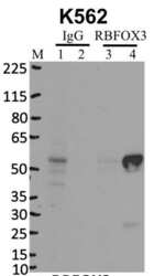
- Immunoprecipitation of RBFOX3 was performed on K562 cells. Antigen-antibody complexes were formed by incubating approximately 500 µg whole cell lysate with 5 µg of RBFOX3 monoclonal antibody (Product # 702022) rotating 60 min at RT. The immune complexes were captured on 625 µg of anti- rabbit coated Dynabeads (Product # 11204D), washed extensively, and eluted with NuPAGE™ LDS Sample Buffer (Product # NP0007). Samples were resolved onto NuPAGE™ 4-12% Bis-Tris gel (Product # NP0335BOX). Lanes 1 and 3 are input and lanes 2 and 4 are IP. Proteins were transferred to PVDF membrane (Product # IB23001). Membrane was blocked in 5% milk. Target was detected using a RBFOX3 monoclonal antibody (Product # 702022) at a dilution of 1:2000, followed by a 1:4000 dilution of secondary antibody. Chemiluminescent detection was performed using ECL Western Blotting Substrate (Product # 32106). Data courtesy of the Yeo lab as part of the ENCODE project (www.encodeproject.org).

Supportive validation
- Submitted by
- Invitrogen Antibodies (provider)
- Main image
- Experimental details
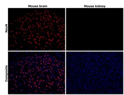
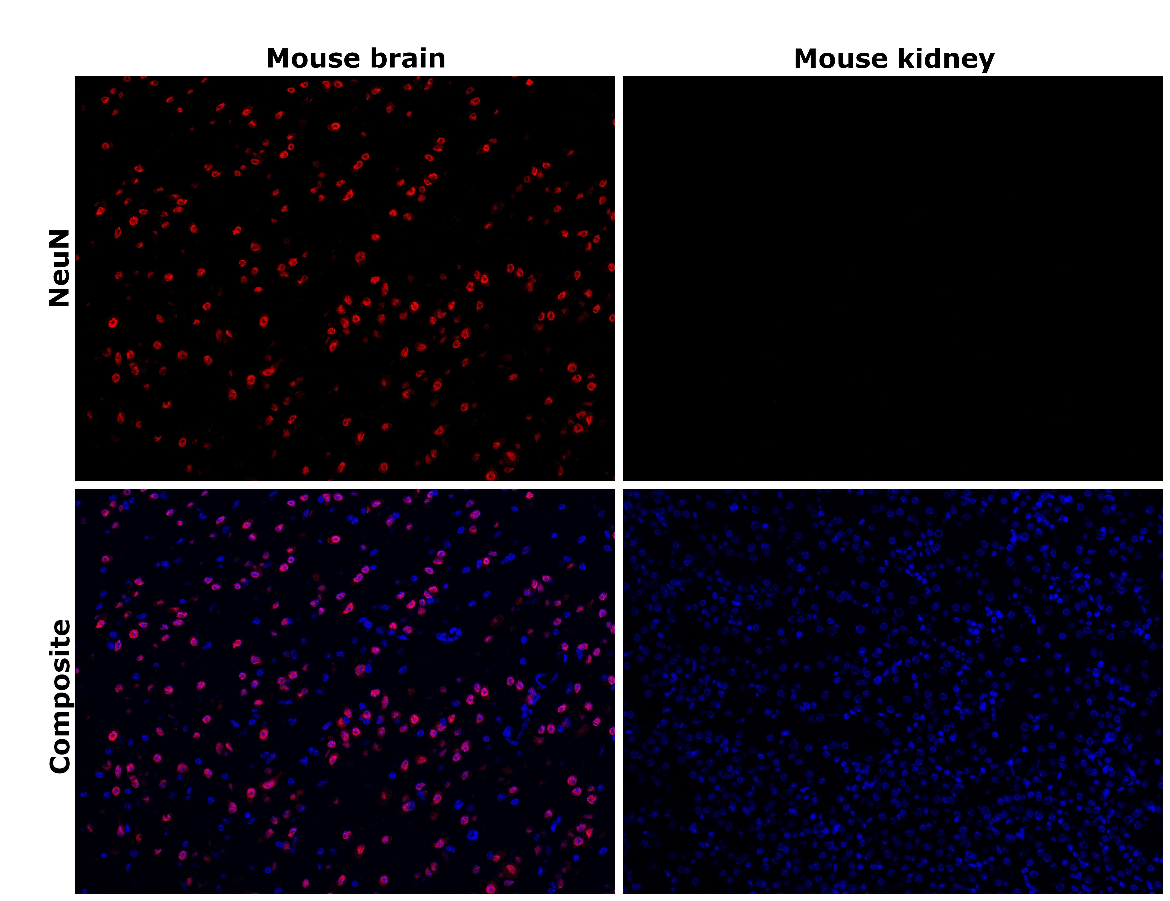
- Immunohistochemical analysis of NeuN was performed using formalin-fixed paraffin-embedded mouse brain and mouse kidney tissue sections. To expose the target protein, heat-induced epitope retrieval was performed on de-paraffinized sections using eBioscience™ IHC Antigen Retrieval Solution - High pH (10X) (Product # 00-4956-58) diluted to 1X solution in water in a decloaking chamber at 110 degree Celsius for 15 minutes. Following antigen retrieval, the sections were blocked with 2% normal goat serum in 1X PBS for 45 minutes at room temperature and then probed with NeuN Recombinant Rabbit Monoclonal Antibody (14H6L24) (Product # 702022) at 5 µg/mL in 0.1% normal goat serum overnight at 4 degree Celsius in a humidified chamber. Detection was performed using Goat anti-Rabbit IgG (H+L) Cross-Adsorbed Secondary Antibody, Alexa Fluor™ 594 (Product # A-11012) at a dilution of 1:2,000 in 0.1% normal goat serum for 45 minutes at room temperature. ReadyProbes™ Tissue Autofluorescence Quenching Kit (Product # R37630) was used to quench autofluorescence from the tissues. Nuclei were stained with DAPI (Product # D1306) and the sections were mounted using ProLong™ Glass Antifade Mountant (Product # P36984). The images were captured on EVOS™ M7000 Imaging System (Product # AMF7000) at 20X magnification and externally deconvoluted. Nuclear expression of NeuN was seen in mouse brain, but not in mouse kidney.
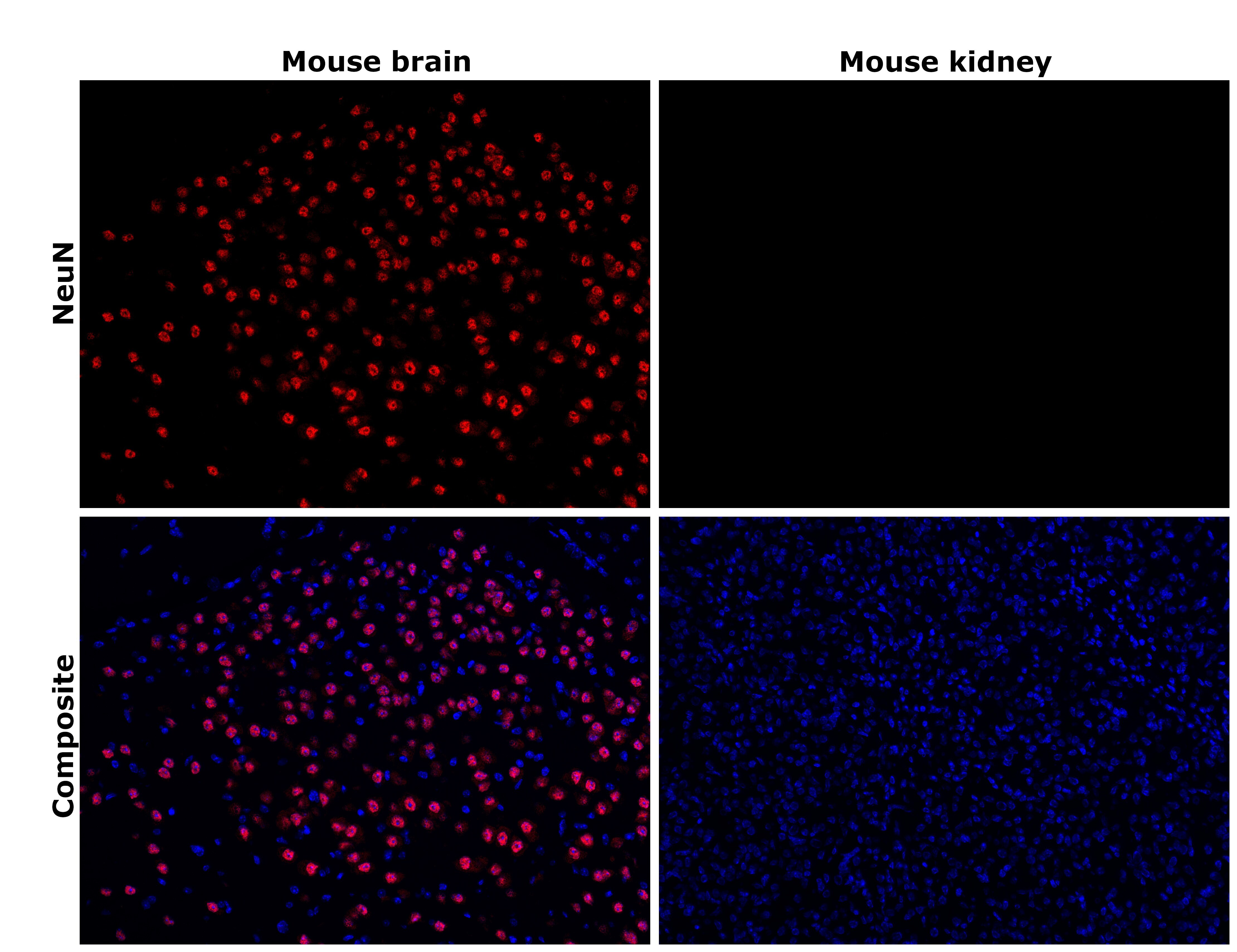
- Submitted by
- Invitrogen Antibodies (provider)
- Main image
- Experimental details
- Immunohistochemical analysis of NeuN was performed using formalin-fixed paraffin-embedded mouse brain and mouse kidney tissue sections. To expose the target protein, heat-induced epitope retrieval was performed on de-paraffinized sections using eBioscience™ IHC Antigen Retrieval Solution - Low pH (10X) (Product # 00-4955-58) diluted to 1X solution in water in a decloaking chamber at 110 degree Celsius for 15 minutes. Following antigen retrieval, the sections were blocked with 2% normal goat serum in 1X PBS for 45 minutes at room temperature and then probed with NeuN Recombinant Rabbit Monoclonal Antibody (14H6L24) (Product # 702022) at 5 µg/mL in 0.1% normal goat serum overnight at 4 degree Celsius in a humidified chamber. Detection was performed using Goat anti-Rabbit IgG (H+L) Cross-Adsorbed Secondary Antibody, Alexa Fluor™ 594 (Product # A-11012) at a dilution of 1:2,000 in 0.1% normal goat serum for 45 minutes at room temperature. ReadyProbes™ Tissue Autofluorescence Quenching Kit (Product # R37630) was used to quench autofluorescence from the tissues. Nuclei were stained with DAPI (Product # D1306) and the sections were mounted using ProLong™ Glass Antifade Mountant (Product # P36984). The images were captured on EVOS™ M7000 Imaging System (Product # AMF7000) at 20X magnification and externally deconvoluted. Nuclear expression of NeuN was seen in mouse brain, but not in mouse kidney.
Supportive validation
- Submitted by
- Invitrogen Antibodies (provider)
- Main image
- Experimental details
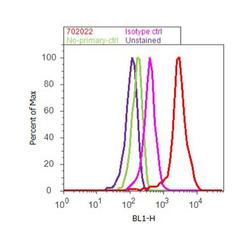
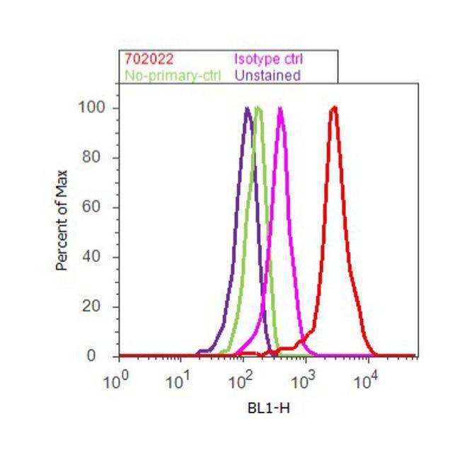
- Flow Cytometry analysis of Fox3 was performed on SH-SY5Y cells labeled with ABfinity™ Anti-Fox3 Recombinant Rabbit Monoclonal Antibody (Product# 702022, 5 ug/ 1M cells) or with rabbit isotype control at 0.5 ug/ml and detected with Goat anti-Rabbit IgG (H+L) Superclonal™ Secondary Antibody, (Alexa Fluor® 488 conjugate, Product # A27034, 0.4 ug/ml, 1:2500) as represented by the red and pink histograms respectively. The purple histogram represents unstained control cells and the green histogram represents no-primary-antibody control. A representative of 10,000 cells were acquired and analyzed for each sample using an Attune® Acoustic Focusing Cytometer (4468770).
Supportive validation
- Submitted by
- Invitrogen Antibodies (provider)
- Main image
- Experimental details
- RNA immunoprecipitation (RIP) western of RBFOX3 was performed on K562 cells. Antigen-antibody complexes were formed by incubating approximately 500 µg whole cell lysate with 5 µg of RBFOX3 monoclonal antibody (Product # 702022) rotating 60 min at RT. The immune complexes were captured on 625 µg of anti- rabbit coated Dynabeads (Product # 11204D), washed extensively, and eluted with NuPAGE™ LDS Sample Buffer (Product # NP0007). Samples were resolved onto NuPAGE™ 4-12% Bis-Tris gel (Product # NP0335BOX). Lanes 1 and 3 are input and lanes 2 and 4 are IP. Proteins were transferred to PVDF membrane (Product # IB23001). Membrane was blocked in 5% milk. Target was detected using a RBFOX3 monoclonal antibody (Product # 702022) at a dilution of 1:2000, followed by a 1:4000 dilution of secondary antibody. Chemiluminescent detection was performed using ECL Western Blotting Substrate (Product # 32106). Data courtesy of the Yeo lab as part of the ENCODE project (www.encodeproject.org).

- Submitted by
- Invitrogen Antibodies (provider)
- Main image
- Experimental details
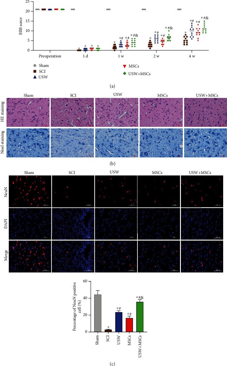
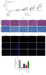
- Figure 1 Combination therapy improves motor functions and reduces spinal cord injury in SCI rats. (a) Evaluated BBB scores in each group of rats in different time-points (preoperation, 1 d, 1 w, 2 w, and 4 w). (b) Histopathological changes in SCI rats treated with monotherapies of USW and HUC-MSCs, respectively, and combination therapy were observed with HE and Nissl staining (scale bar = 50 mu m). (c) Expression level of NeuN detected with IF (scale bar = 50 mu m); percentage of NeuN positive cells was calculated and represented in bar chart attached. "" * , #, &"" indicated significant difference ( p < 0.05), "" * "" vs. Sham group, ""#"" vs. SCI group, and ""&"" vs. rats treated with monotherapy (USW/HUC-MSCs).