Explore
 Validate
 Learn
Learn Western blot
Antibody data
- Antibody Data
- Antigen structure
- References [2]
- Comments [0]
- Validations
- Western blot [1]
- Immunohistochemistry [1]
- Flow cytometry [1]
Submit
Validation data
Reference
Comment
Report error
- Product number
- ABIN453367 - Provider product page

- Provider
- antibodies-online
- Product name
- anti-serpin Peptidase Inhibitor, Clade D (Heparin Cofactor), Member 1 (SERPIND1) (AA 226-256), (Middle Region) antibody
- Antibody type
- Polyclonal
- Antigen
- KLH conjugated synthetic peptide selected from aa 226-256 of the Center region of Human SERPIND1 Genename: SERPIND1
- Description
- Saturated Ammonium Sulfate (SAS) Precipitation
- Reactivity
- Human
- Host
- Rabbit
- Epitope
- AA 226-256,Middle Region
- Vial size
- 0.4 mL
- Storage
- Store the antibody undiluted at 2-8°C for one month or (in aliquots) at -20°C for longer.
- Handling
- Avoid repeated freezing and thawing.
Submitted references Plasma heparin cofactor II activity is an independent predictor of future cardiovascular events in patients after acute myocardial infarction.
Placental dermatan sulfate: isolation, anticoagulant activity, and association with heparin cofactor II.
Huang SS, Huang PH, Chen YH, Sung SH, Chiang KH, Chen JW, Lin SJ
Coronary artery disease 2008 Dec;19(8):597-602
Coronary artery disease 2008 Dec;19(8):597-602
Placental dermatan sulfate: isolation, anticoagulant activity, and association with heparin cofactor II.
Giri TK, Tollefsen DM
Blood 2006 Apr 1;107(7):2753-8
Blood 2006 Apr 1;107(7):2753-8
No comments: Submit comment
Supportive validation
- Submitted by
- antibodies-online (provider)
- Main image
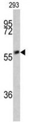
- Experimental details
- Western blot analysis of SERPIND1 Antibody (Center) (AP17736PU-N) in 293 cell line lysates (35ug/lane). SERPIND1 (arrow) was detected using the purified Pab.

Supportive validation
- Submitted by
- antibodies-online (provider)
- Main image
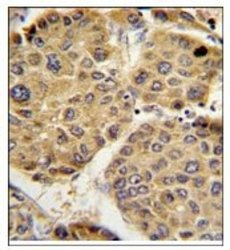
- Experimental details
- AP17736PU-N SERPIND1 Antibody staining of Formalin-Fixed, Paraffin-Embedded Human hepatocarcinoma using Peroxidase-conjugated secondary antibody, followed by DAB staining. This data demonstrates the use of this antibody for immunohistochemistry. Clinical relevance has not been evaluated.

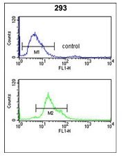
Supportive validation
- Submitted by
- antibodies-online (provider)
- Main image

- Experimental details
- Flow Cytometry analysis of 293 cells (bottom histogram) compared to a Negative Control cell (top histogram) using AP17736PU-N SERPIND1 Antibody. FITC-conjugated goat-anti-rabbit secondary antibodies were used for the analysis.