Explore
 Validate
 Learn
Learn Western blot
 Immunohistochemistry
Antibody data
- Antibody Data
- Antigen structure
- References [2]
- Comments [0]
- Validations
- Immunohistochemistry [1]
- Other assay [4]
Submit
Validation data
Reference
Comment
Report error
- Product number
- PA5-30996 - Provider product page

- Provider
- Invitrogen Antibodies
- Product name
- CBX2 Polyclonal Antibody
- Antibody type
- Polyclonal
- Antigen
- Recombinant full-length protein
- Description
- Recommended positive controls: A431, Jurkat. Store product as a concentrated solution. Centrifuge briefly prior to opening the vial.
- Reactivity
- Human, Mouse
- Host
- Rabbit
- Isotype
- IgG
- Vial size
- 100 μL
- Concentration
- 0.9 mg/mL
- Storage
- Store at 4°C short term. For long term storage, store at -20°C, avoiding freeze/thaw cycles.
Submitted references CBX2 identified as driver of anoikis escape and dissemination in high grade serous ovarian cancer.
Identification of the epigenetic reader CBX2 as a potential drug target in advanced prostate cancer.
Wheeler LJ, Watson ZL, Qamar L, Yamamoto TM, Post MD, Berning AA, Spillman MA, Behbakht K, Bitler BG
Oncogenesis 2018 Nov 26;7(11):92
Oncogenesis 2018 Nov 26;7(11):92
Identification of the epigenetic reader CBX2 as a potential drug target in advanced prostate cancer.
Clermont PL, Crea F, Chiang YT, Lin D, Zhang A, Wang JZ, Parolia A, Wu R, Xue H, Wang Y, Ding J, Thu KL, Lam WL, Shah SP, Collins CC, Wang Y, Helgason CD
Clinical epigenetics 2016;8:16
Clinical epigenetics 2016;8:16
No comments: Submit comment
Supportive validation
- Submitted by
- Invitrogen Antibodies (provider)
- Main image
- Experimental details
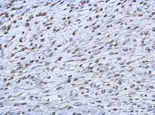
- Immunohistochemical analysis of paraffin-embedded C2C12 xenograft, using CBX2 (Product # PA5-30996) antibody at 1:500 dilution. Antigen Retrieval: EDTA based buffer, pH 8.0, 15 min.
Supportive validation
- Submitted by
- Invitrogen Antibodies (provider)
- Main image
- Experimental details
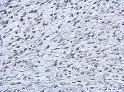
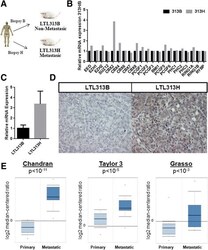
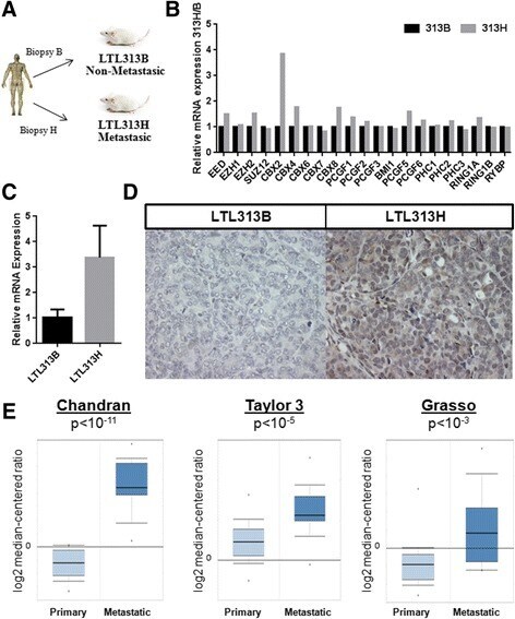
- Fig. 1 CBX2 is overexpressed in metastatic PCa. a Establishment of the LTL313B/LTL313H PDX model of metastatic PCa; b Expression of core PcG family members in the LTL313H/LTL313B xenograft model; Results are based on a single microarray experiment; c Confirmation of CBX2 mRNA up-regulation in the LTL313H tumor line by qRT-PCR; d Confirmation of CBX2 protein up-regulation in the LTL313H tumor line by IHC (20x). Images are representative of multiple fields taken from 2 independent experiments; e Elevated CBX2 mRNA levels in metastatic PCa compared to non-metastatic samples in three independent patients
- Submitted by
- Invitrogen Antibodies (provider)
- Main image

- Experimental details
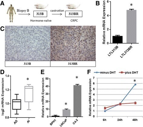
- Fig. 2 Hormonal regulation of CBX2. a Establishment of the LTL313B/LTL313BR patient-derived xenograft model of CRPC; b Assessment of CBX2 mRNA levels in the LTL313B/LTL313BR xenograft model by qRT-PCR; c IHC staining of CBX2 in the LTL313B and LTL313BR xenografts x20. Images are representative of multiple fields taken from two independent experiments; d Levels of CBX2 mRNA in androgen-dependent (AD, n = 10) and androgen-independent (AI, n = 5) PDXs from the LTL; e Relative CBX2 expression in PCa cell lines compared to benign control (BPH1) assessed by qRT-PCR; f CBX2 mRNA levels in LNCaP cells cultured in charcoaled-stripped media in the presence or absence of DHT supplementation (10 nM)
- Submitted by
- Invitrogen Antibodies (provider)
- Main image

- Experimental details
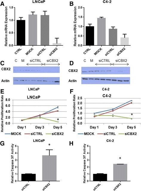
- Fig. 3 CBX2 depletion induces proliferation arrest and apoptosis in advanced PCa cell lines. a , b Confirmation of CBX2 mRNA knockdown in LNCaP and C4-2 cells by qPCR; c , d Confirmation of CBX2 protein knockdown in LNCaP and C4-2 cells; e , f MTT analysis of cell viability following CBX2 silencing in LNCaP and C4-2 cells; g , h Assessment of caspase 3-7 activity in LNCaP and C4-2 cells following CBX2 depletion
- Submitted by
- Invitrogen Antibodies (provider)
- Main image

- Experimental details
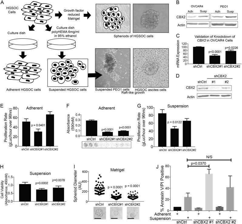
- Fig. 2 Inhibition of CBX2 impairs HGSOC cell proliferation. a Model describing basic protocol for establishing adherent, suspension, and spheroid growth environments. For adherent and suspension, two verified high grade serous ovarian carcinoma cell lines were initially grown on tissue culture plastic, then distributed to normal tissue culture dishes (adherent) and polyHEMA coated culture dish (suspension) growth environments. Distributed at 1:3 or 1:5 ratio to account for forced suspension induced cell death. Photographs show PEO1 cells after 7 days in suspension (left) and HGSOC cells directly derived from patient ascites. For spheroid formation, cells were grown in 3D in Matrigel for 12 days. A representative image of a resulting spheroid is shown at upper right. b Immunoblots against CBX2 protein from OVCAR4, and PEO1 cells grown in adherent and suspension settings (described in ( a )) over 7 days. c RT-qPCR for CBX2 in OVCAR4 cells transduced with small hairpin RNA (shRNA) specific for CBX2 . Representative mRNA expression of CBX2 in shControl, shCBX2#1, and shCBX2#2. Statistical test = ANOVA. d Immunoblots against CBX2 protein derived from OVCAR4 shControl, shCBX2#1, and shCBX2#2 transduced cells (beta-actin = loading control). e Proliferation assay of OVCAR4 cells with CBX2 knockdown and shControl, grown in adherent setting (tissue culture plastic) over 96 h, evaluated using gLuc activity. Statistical test = ANOVA. f Same as ( e ), crystal violet staini