Explore
 Validate
 Learn
Learn Western blot
 Immunohistochemistry
Antibody data
- Antibody Data
- Antigen structure
- References [1]
- Comments [0]
- Validations
- Immunohistochemistry [1]
- Flow cytometry [2]
- Other assay [1]
Submit
Validation data
Reference
Comment
Report error
- Product number
- PA5-14361 - Provider product page

- Provider
- Invitrogen Antibodies
- Product name
- Anti-IGHA1
- Antibody type
- Polyclonal
- Antigen
- Synthetic peptide from the C-terminal region of IGHA1 conjugated to KLH.
- Reactivity
- Human
- Host
- Rabbit
- Vial size
- 100 ug
- Concentration
- 0.25 mg/ml
- Storage
- Maintain refrigerated at 2-8°C for up to 6 months. For long term storage store at -20°C
Submitted references Salivary Proteome Changes in Response to Acute Psychological Stress Due to an Oral Exam Simulation in University Students: Effect of an Olfactory Stimulus.
Zallocco L, Giusti L, Ronci M, Mussini A, Trerotola M, Mazzoni MR, Lucacchini A, Sebastiani L
International journal of molecular sciences 2021 Apr 21;22(9)
International journal of molecular sciences 2021 Apr 21;22(9)
No comments: Submit comment
Supportive validation
- Submitted by
- Invitrogen Antibodies (provider)
- Main image

- Experimental details
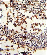
- Immunohistochemistry analysis of IGHA1 in formalin-fixed and paraffin-embedded human lymph. Samples were incubated with IGHA1 polyclonal antibody (Product # PA5-14361) which was peroxidase-conjugated to the secondary antibody, followed by DAB staining. This data demonstrates the use of this antibody for immunohistochemistry; clinical relevance has not been evaluated.
Supportive validation
- Submitted by
- Invitrogen Antibodies (provider)
- Main image
- Experimental details
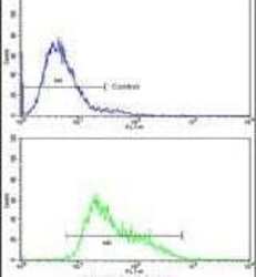
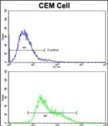
- Flow cytometry analysis of CEM cells using an IGHA1 polyclonal antibody (Product # PA5-14361) (bottom), compared to a negative control cell (top) at a dilution of 1:10-50, followed by a FITC-conjugated goat anti-rabbit antibody
- Submitted by
- Invitrogen Antibodies (provider)
- Main image
- Experimental details
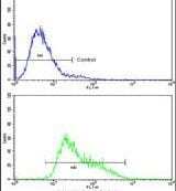
- Flow cytometry of IGHA1 in CEM cells (bottom histogram). Samples were incubated with IGHA1 polyclonal antibody (Product # PA5-14361) followed by FITC-conjugated goat-anti-rabbit secondary antibody. Negative control cell (top histogram).

Supportive validation
- Submitted by
- Invitrogen Antibodies (provider)
- Main image
- Experimental details
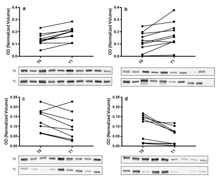
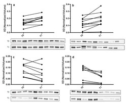
- Figure 3 Validation of alpha-amylase (panel ( a , b )) and immunoglobulin alpha chain (IGHA) (panel ( c , d )) in whole saliva (WS) samples at T0 (after relaxation phase) and T1 (after anxiety test) from control (panel ( a , c )) and odor exposed (panel ( b , d )) subjects using Western blot (WB) analysis. A graphical representation of normalized optical density (OD) of alpha-amylase and IGHA bands is shown. Each pair of connected points represents one experiment using T0 and T1 WS samples obtained from a single subject. The p -values were determined by paired t-test. Representative blots are shown below the graphs. A single immunoreactive band with apparent molecular weight approximately of 55 kDa and 67 kDa was obtained for alpha-amylase and IGHA, respectively.