 Explore
 Validate
 Learn
Learn Western blot
Antibody data
- Antibody Data
- Antigen structure
- References [0]
- Comments [0]
- Validations
- Western blot [1]
- Immunocytochemistry [3]
- Immunohistochemistry [1]
Submit
Validation data
Reference
Comment
Report error
- Product number
- NBP2-29961 - Provider product page

- Provider
- Novus Biologicals
- Product name
- Mouse Monoclonal Lactoferrin Antibody
- Antibody type
- Monoclonal
- Description
- Protein G purified.
- Reactivity
- Human
- Host
- Mouse
- Isotype
- IgG
- Vial size
- 0.4 ml
- Storage
- Store at 4C short term. Aliquot and store at -20C long term. Avoid freeze-thaw cycles.
No comments: Submit comment
Supportive validation
- Submitted by
- Novus Biologicals (provider)
- Main image

- Experimental details
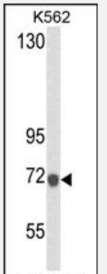
- Western Blot: Lactoferrin Antibody (119CT80.1.1) [NBP2-29961] - Western blot analysis of LTF (NBP2-29961) in K562 Cell Line lysates (35ug/lane). LTF (arrow) was detected using the purified Mab.(1ug/ml)
Supportive validation
- Submitted by
- Novus Biologicals (provider)
- Main image
- Experimental details
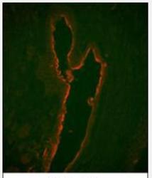
- Immunocytochemistry/Immunofluorescence: Lactoferrin Antibody (119CT80.1.1) [NBP2-29961] - Immunofluorescence analysis of LTF Monoclonal Antibody with paraffin-embedded human prostate carcinoma tissue . 0.05 mg/ml primary antibody was followed by PE-conjugated goat anti-mouse lgG (whole molecule). PE emits orange fluorescence.
- Submitted by
- Novus Biologicals (provider)
- Main image

- Experimental details
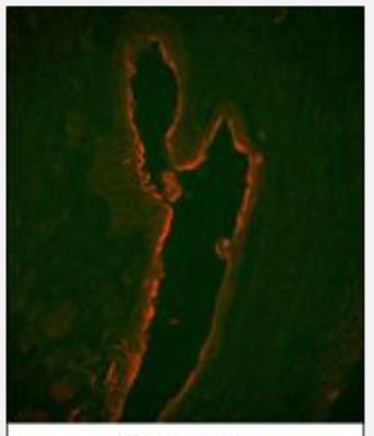
- Immunocytochemistry/Immunofluorescence: Lactoferrin Antibody (119CT80.1.1) [NBP2-29961] - Confocal immunofluorescent analysis of LTF Antibody (NBP2-29961) with HepG2 cell followed by Alexa Fluor 488-conjugated goat anti-mouse lgG (green). DAPI was used to stain the cell nuclear (blue).
- Submitted by
- Novus Biologicals (provider)
- Main image

- Experimental details
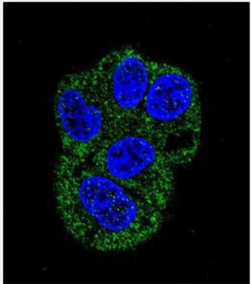
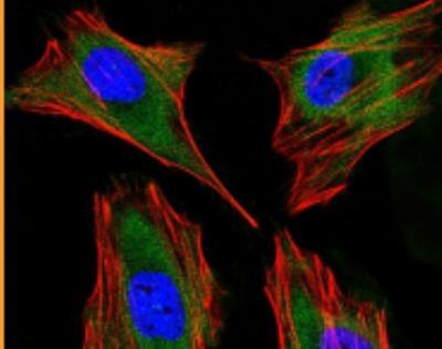
- Immunocytochemistry/Immunofluorescence: Lactoferrin Antibody (119CT80.1.1) [NBP2-29961] - Immunofluorescent analysis of 4% paraformaldehyde-fixed, 0. 1% Triton X-100 permeabilized Hela (Human Cervical epithelial adenocarcinoma cell line) cells labeling Pdx1 with AM1819a at 1/25 dilution, followed by Dylight (R) 488-conjugated goat anti-mouse IgG (NA166821) secondary antibody at 1/200 dilution (green). Immunofluorescence image showing cytoplasm staining on Hela cell line. Cytoplasmic actin is detected with Dylight (R) 554 Phalloidin at 1/100 dilution (red). The nuclear counter stain is DAPI (blue).
Supportive validation
- Submitted by
- Novus Biologicals (provider)
- Main image

- Experimental details
- Immunohistochemistry-Paraffin: Lactoferrin Antibody (119CT80.1.1) [NBP2-29961] - Formalin-fixed and paraffin-embedded human prostate carcinoma with LTF Monoclonal Antibody, which was peroxidase-conjugated to the secondary antibody, followed by DAB staining. This data demonstrates the use of this antibody for immunohistochemistry; clinical relevance has not been evaluated.
