Explore
 Validate
 Learn
Learn Western blot
Antibody data
- Antibody Data
- Antigen structure
- References [2]
- Comments [0]
- Validations
- Western blot [1]
Submit
Validation data
Reference
Comment
Report error
- Product number
- AF6610 - Provider product page

- Provider
- R&D Systems
- Product name
- Human/Mouse Peroxiredoxin 3 Antibody
- Antibody type
- Polyclonal
- Description
- Immunogen affinity purified. Detects human and mouse Peroxiredoxin 3 in direct ELISAs and Western blots. In direct ELISAs, approximately 6% cross-reactivity with recombinant mouse (rm) Peroxiredoxin 2 is observed and less than 1% cross-reactivity with recombinant human (rh) Peroxiredoxin 1, rmPeroxiredoxin 5, and rmPeroxiredoxin 6 is observed.
- Reactivity
- Human, Mouse
- Host
- Goat
- Conjugate
- Unconjugated
- Antigen sequence
P20108- Isotype
- IgG
- Vial size
- 100 ug
- Concentration
- LYOPH
- Storage
- Use a manual defrost freezer and avoid repeated freeze-thaw cycles. 12 months from date of receipt, -20 to -70 °C as supplied. 1 month, 2 to 8 °C under sterile conditions after reconstitution. 6 months, -20 to -70 °C under sterile conditions after reconstitution.
Submitted references Electrophiles modulate glutathione reductase activity via alkylation and upregulation of glutathione biosynthesis.
NOX2 amplifies acetaldehyde-mediated cardiomyocyte mitochondrial dysfunction in alcoholic cardiomyopathy.
Jobbagy S, Vitturi DA, Salvatore SR, Turell L, Pires MF, Kansanen E, Batthyany C, Lancaster JR Jr, Freeman BA, Schopfer FJ
Redox biology 2019 Feb;21:101050
Redox biology 2019 Feb;21:101050
NOX2 amplifies acetaldehyde-mediated cardiomyocyte mitochondrial dysfunction in alcoholic cardiomyopathy.
Brandt M, Garlapati V, Oelze M, Sotiriou E, Knorr M, Kröller-Schön S, Kossmann S, Schönfelder T, Morawietz H, Schulz E, Schultheiss HP, Daiber A, Münzel T, Wenzel P
Scientific reports 2016 Sep 14;6:32554
Scientific reports 2016 Sep 14;6:32554
No comments: Submit comment
Supportive validation
- Submitted by
- R&D Systems (provider)
- Main image
- Experimental details
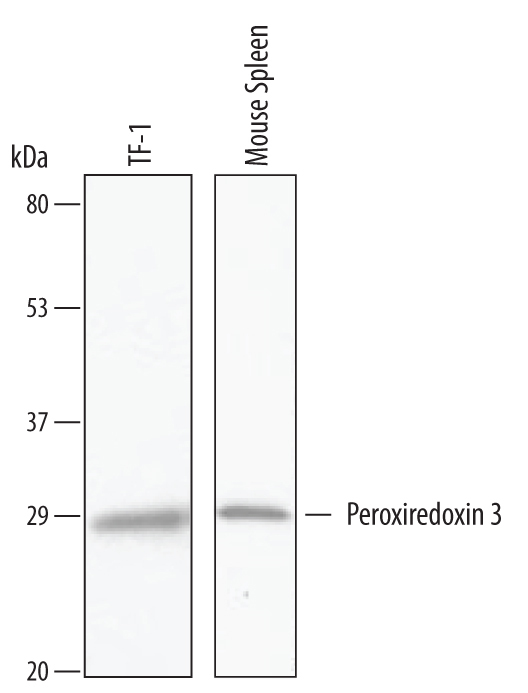
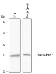
- Detection of Human and Mouse Peroxiredoxin 3 by Western Blot. Western blot shows lysates of TF-1 human erythroleukemic cell line and mouse spleen tissue. PVDF Membrane was probed with 1 µg/mL of Human/Mouse Peroxiredoxin 3 Antigen Affinity-purified Polyclonal Antibody (Catalog # AF6610) followed by HRP-conjugated Anti-Goat IgG Secondary Antibody (Catalog # HAF019). A specific band was detected for Peroxiredoxin 3 at approximately 28 kDa (as indicated). This experiment was conducted under reducing conditions and using Immunoblot Buffer Group 8.