Explore
 Validate
 Learn
Learn Western blot
Antibody data
- Antibody Data
- Antigen structure
- References [1]
- Comments [0]
- Validations
- Western blot [1]
- Immunohistochemistry [1]
Submit
Validation data
Reference
Comment
Report error
- Product number
- AF5460 - Provider product page

- Provider
- Novus Biologicals
- Product name
- Goat Polyclonal Peroxiredoxin 4 Antibody
- Antibody type
- Polyclonal
- Description
- Antigen Affinity-purified. Detects human, mouse and rat Peroxiredoxin 4 in Western blots.
- Reactivity
- Human, Mouse, Rat
- Host
- Goat
- Conjugate
- Unconjugated
- Isotype
- IgG
- Vial size
- 100 ug
- Concentration
- LYOPH
- Storage
- Use a manual defrost freezer and avoid repeated freeze-thaw cycles. 12 months from date of receipt, -20 to -70 degreesC as supplied. 1 month, 2 to 8 degreesC under sterile conditions after reconstitution. 6 months, -20 to -70 degreesC under sterile conditions after reconstitution.
Submitted references 1,25-Dihydroxy vitamin D prevents tumorigenesis by inhibiting oxidative stress and inducing tumor cellular senescence in mice.
Chen L, Yang R, Qiao W, Yuan X, Wang S, Goltzman D, Miao D
International journal of cancer 2018 Jul 15;143(2):368-382
International journal of cancer 2018 Jul 15;143(2):368-382
No comments: Submit comment
Supportive validation
- Submitted by
- Novus Biologicals (provider)
- Main image
- Experimental details
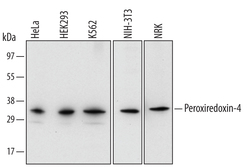
- Detection of Human/Mouse/Rat Peroxiredoxin 4 by Western Blot. Western blot shows lysates of HeLa human cervical epithelial carcinoma cell line, HEK293 human embryonic kidney cell line, K562 human chronic myelogenous leukemia cell line, NIH-3T3 mouse embryonic fibroblast cell line, and NRK rat normal kidney cell line. PVDF membrane was probed with 1 µg/mL of Goat Anti-Human/Mouse/Rat Peroxiredoxin 4 Antigen Affinity-purified Polyclonal Antibody (Catalog # AF5460) followed by HRP-conjugated Anti-Goat IgG Secondary Antibody (Catalog # HAF109). A specific band was detected for Peroxiredoxin 4 at approximately 32 kDa (as indicated). This experiment was conducted under reducing conditions and using Immunoblot Buffer Group 2.

Supportive validation
- Submitted by
- Novus Biologicals (provider)
- Main image
- Experimental details
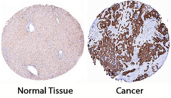
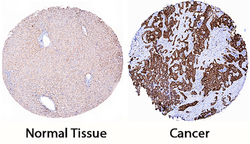
- Peroxiredoxin 4 in Human Liver Cancer Tissue. Peroxiredoxin 4 was detected in immersion fixed paraffin-embedded sections of human liver cancer tissue using Goat Anti-Human/Mouse/Rat Peroxiredoxin 4 Antigen Affinity-purified Polyclonal Antibody (Catalog # AF5460) at 0.3 µg/mL for 1 hour at room temperature followed by incubation with the Anti-Goat IgG VisUCyte™ HRP Polymer Antibody (Catalog # VC004). Tissue was stained using DAB (brown) and counterstained with hematoxylin (blue). Specific staining was localized to cancer cells. View our protocol for IHC Staining with VisUCyte HRP Polymer Detection Reagents.