Explore
 Validate
 Learn
Learn Western blot
 Immunocytochemistry
 Immunohistochemistry
Antibody data
- Antibody Data
- Antigen structure
- References [4]
- Comments [0]
- Validations
- Immunocytochemistry [2]
- Other assay [2]
Submit
Validation data
Reference
Comment
Report error
- Product number
- PA3-753 - Provider product page

- Provider
- Invitrogen Antibodies
- Product name
- PRDX4 Polyclonal Antibody
- Antibody type
- Polyclonal
- Antigen
- Purifed from natural sources
- Description
- PA3-753 detects Peroxiredoxin 4 protein in human samples. This antibody is specific for Prx 4 and shows no cross-reactivity with other Prx isoforms. PA3-753 has successfully been used in Western blot and immunocytochemistry procedures. By Western blot, this antibody detects an ~23 kDa and ~20 kDa protein representing pro-Prx 4 and mature Prx 4, respectively, in human PC3 cell lysate. The PA3-753 antigen is purified human recombinant Prx 4 from K562 cells.
- Reactivity
- Human
- Host
- Rabbit
- Isotype
- IgG
- Vial size
- 100 µL
- Concentration
- Conc. Not Determined
- Storage
- -20° C, Avoid Freeze/Thaw Cycles
Submitted references Overexpression of PRDX4 Modulates Tumor Microenvironment and Promotes Urethane-Induced Lung Tumorigenesis.
Peroxiredoxin 4 promotes embryonal hepatoblastoma cell migration but induces fetal cell differentiation.
Quantitative Proteomics Reveal Peroxiredoxin Perturbation Upon Persistent Lymphocytic Choriomeningitis Virus Infection in Human Cells.
Nonredundant antioxidant defense by multiple two-cysteine peroxiredoxins in human prostate cancer cells.
Zheng J, Guo X, Nakamura Y, Zhou X, Yamaguchi R, Zhang J, Ishigaki Y, Uramoto H, Yamada S
Oxidative medicine and cellular longevity 2020;2020:8262730
Oxidative medicine and cellular longevity 2020;2020:8262730
Peroxiredoxin 4 promotes embryonal hepatoblastoma cell migration but induces fetal cell differentiation.
Zheng J, Guo X, Shioya A, Yoshioka T, Matsumoto K, Hiraki T, Kusano H, Oyama T, Kurose N, Yamaguchi R, Uramoto H, Ieiri S, Okajima H, Kohno M, Yamada S
American journal of translational research 2020;12(6):2726-2737
American journal of translational research 2020;12(6):2726-2737
Quantitative Proteomics Reveal Peroxiredoxin Perturbation Upon Persistent Lymphocytic Choriomeningitis Virus Infection in Human Cells.
Benej M, Danchenko M, Oveckova I, Cervenak F, Tomaska L, Grossmannova K, Polcicova K, Golias T, Tomaskova J
Frontiers in microbiology 2019;10:2438
Frontiers in microbiology 2019;10:2438
Nonredundant antioxidant defense by multiple two-cysteine peroxiredoxins in human prostate cancer cells.
Shen C, Nathan C
Molecular medicine (Cambridge, Mass.) 2002 Feb;8(2):95-102
Molecular medicine (Cambridge, Mass.) 2002 Feb;8(2):95-102
No comments: Submit comment
Supportive validation
- Submitted by
- Invitrogen Antibodies (provider)
- Main image
- Experimental details
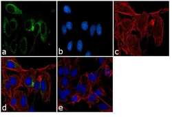
- Immunofluorescence analysis of Peroxiredoxin 4 was performed using 70% confluent log phase LNCaP cells. The cells were fixed with 4% paraformaldehyde for 10 minutes, permeabilized with 0.1% Triton™ X-100 for 10 minutes, and blocked with 1% BSA for 1 hour at room temperature. The cells were labeled with PRDX4 Rabbit Polyclonal Antibody (Product # PA3-753) at 1:250 dilution in 0.1% BSA and incubated for 3 hours at room temperature and then labeled with Goat anti-Rabbit IgG (H+L) Superclonal™ Secondary Antibody, Alexa Fluor® 488 conjugate (Product # A27034) at a dilution of 1:2000 for 45 minutes at room temperature (Panel a: green). Nuclei (Panel b: blue) were stained with SlowFade® Gold Antifade Mountant with DAPI (Product # S36938). F-actin (Panel c: red) was stained with Rhodamine Phalloidin (Product # R415, 1:300). Panel d represents the merged image showing cytoplasmic localization. Panel e shows the no primary antibody control. The images were captured at 60X magnification.

- Submitted by
- Invitrogen Antibodies (provider)
- Main image
- Experimental details
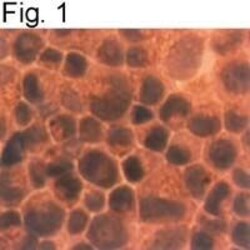
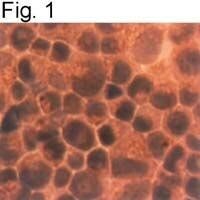
- Immunocytochemical staining of Prx 4 in TSU-Pr1 cells using Product # PA3-753.
Supportive validation
- Submitted by
- Invitrogen Antibodies (provider)
- Main image
- Experimental details
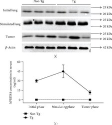
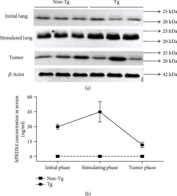
- Figure 1 Elevated expressions of PRDX4 in Tg mice after urethane stimulation. (a) Comparisons of PRDX4 expression between Tg and non-Tg controls in initial lung tissues, in lung tissues after urethane stimulation, and in lung tumor tissues, respectively, by Western blotting. (b) Serum hPRDX4 expressions detected by the ELISA method in the process of tumor formation. The values represent mean +- SD, and the experiments shown were repeated in triplicate with similar results.
- Submitted by
- Invitrogen Antibodies (provider)
- Main image

- Experimental details
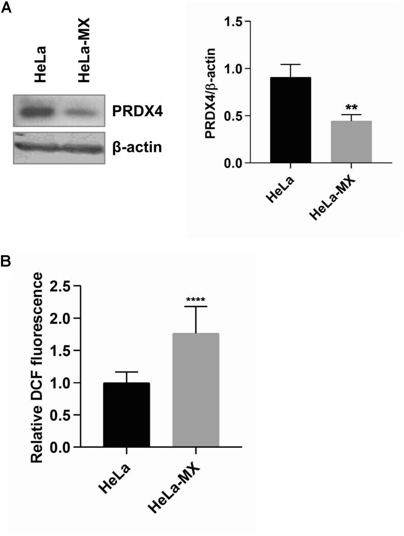
- FIGURE 5 ROS production in LCMV- and mock-infected HeLa cells. (A) Immunoblot analysis of PRDX4 (left) with specific antibody using whole-cell extracts prepared from LCMV-infected HeLa cells (HeLa-MX) and mock-infected HeLa cells (HeLa). The signal obtained with anti-beta-actin antibody was used as loading control. One representative of three biological replicates is shown. Densitometric analysis of protein abundance (right) was performed using ImageJ software. Relative quantity of proteins was calculated as the ratio of the intensity of each signal to the intensity of the related beta-actin internal standard. Values represent the means of three biological replicates. Error bars denote standard deviations. ** P < 0.01 (HeLa-MX vs. HeLa). (B) ROS generation was assessed in a microplate reader using DCF fluorescence and normalized to the number of cells measured by DAPI staining. The results represent the mean from three independent biological experiments, each done in eight replicates. Error bars denote standard deviations. Data are presented as relative increase compared to control (HeLa cells), which was set to 1. **** P < 0.0001 (HeLa-MX vs. HeLa).