Explore
 Validate
 Learn
Learn ELISA
ELISA Immunocytochemistry
Antibody data
- Antibody Data
- Antigen structure
- References [0]
- Comments [0]
- Validations
- Immunocytochemistry [1]
- Flow cytometry [1]
Submit
Validation data
Reference
Comment
Report error
- Product number
- MA5-48248 - Provider product page

- Provider
- Invitrogen Antibodies
- Product name
- TSPAN1 Chimeric Recombinant Rabbit Monoclonal Antibody (4/12)
- Antibody type
- Monoclonal
- Antigen
- Other
- Description
- Specificity: This antibody is specific for the largest extracellular domain of tetraspanin 1.
- Reactivity
- Human
- Host
- Rabbit
- Isotype
- IgG
- Antibody clone number
- 4/12
- Vial size
- 200 μg
- Concentration
- 1 mg/mL
- Storage
- Store at 4°C short term. For long term storage, store at -20°C, avoiding freeze/thaw cycles.
No comments: Submit comment
Supportive validation
- Submitted by
- Invitrogen Antibodies (provider)
- Main image
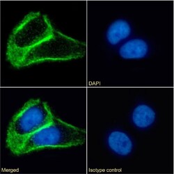
- Experimental details
- Immunocytochemistry/Immunofluorescence analysis of TSPAN1 in A431 cells using TSPAN1 Chimeric Monoclonal Antibody (Product # MA5-48248). Paraformaldehyde-fixed cells were immobilized on cover slips and stained with TSPAN1 Chimeric Monoclonal Antibody at 10 µg/mL for 1h followed by Alexa Fluor 488 secondary antibody (2 µg/mL), showing membrane staining. The nuclear stain is DAPI (blue). Panels show from left-right, top-bottom TSPAN1 Chimeric Monoclonal Antibody, DAPI, merged channels and an isotype control. The isotype control was a Mouse IgG1 Isotype Control Antibody (Product # MA5-47823) followed by staining with Alexa Fluor 488 secondary antibody.

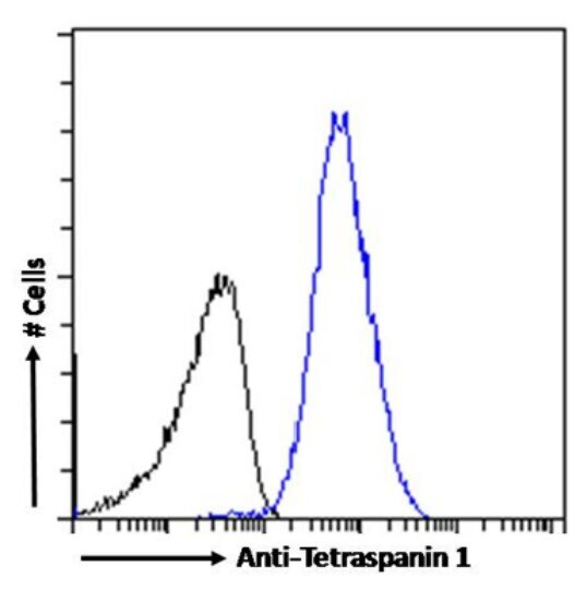
Supportive validation
- Submitted by
- Invitrogen Antibodies (provider)
- Main image

- Experimental details
- Flow Cytometry analysis of TSPAN1 in A431 cells using TSPAN1 Chimeric Monoclonal Antibody (Product # MA5-48248). Cells were fixed with paraformaldehyde, permeabilized with 0.5% triton and stained with Rabbit IgG Isotype Control (Product # MA5-47825) (black line) or TSPAN1 Chimeric Monoclonal Antibody (blue line) at a dilution of 1:100 for 1h at RT. After washing, the bound antibody was detected using a goat anti-rabbit IgG AF488 antibody at a dilution of 1:1,000 and cells analyzed using a flow-cytometer.