Explore
 Validate
 Learn
LearnPA5-98085
antibody from Invitrogen Antibodies
Targeting: PRDX5
ACR1, AOEB166, B166, MGC117264, MGC142283, MGC142285, PLP, PMP20, PRDX6, PRXV, SBBI10
 Western blot
 ELISA
ELISA Immunocytochemistry
Antibody data
- Antibody Data
- Antigen structure
- References [1]
- Comments [0]
- Validations
- Immunocytochemistry [2]
- Immunohistochemistry [3]
- Other assay [2]
Submit
Validation data
Reference
Comment
Report error
- Product number
- PA5-98085 - Provider product page

- Provider
- Invitrogen Antibodies
- Product name
- PRDX5 Polyclonal Antibody
- Antibody type
- Polyclonal
- Antigen
- Recombinant full-length protein
- Reactivity
- Human, Mouse
- Host
- Rabbit
- Isotype
- IgG
- Vial size
- 100 μL
- Concentration
- 0.5 mg/mL
- Storage
- -20°C or -80°C if preferred
Submitted references Expression of antioxidant enzymes in lesions of multiple sclerosis and its models.
Moezzi D, Dong Y, Jain RW, Lozinski BM, Ghorbani S, D'Mello C, Wee Yong V
Scientific reports 2022 Jul 26;12(1):12761
Scientific reports 2022 Jul 26;12(1):12761
No comments: Submit comment
Supportive validation
- Submitted by
- Invitrogen Antibodies (provider)
- Main image
- Experimental details
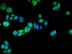
- Immunofluorescent analysis of PRDX5 in PC-3 cells using a PRDX5 polyclonal antibody (Product # PA5-98085) at a dilution of 1:100. Alexa Fluor 488-congugated Goat Anti-Rabbit IgG(H+L) secondary antibody was used.

- Submitted by
- Invitrogen Antibodies (provider)
- Main image

- Experimental details
- Immunofluorescent analysis of PRDX5 in PC-3 cells using a PRDX5 polyclonal antibody (Product # PA5-98085) at a dilution of 1:100. Alexa Fluor 488-congugated Goat Anti-Rabbit IgG(H+L) secondary antibody was used.

Supportive validation
- Submitted by
- Invitrogen Antibodies (provider)
- Main image
- Experimental details
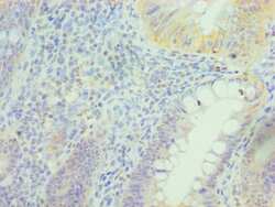
- Immunohistochemical analysis of PRDX5 in paraffin embedded human epityphlon tissue using a PRDX5 polyclonal antibody (Product # PA5-98085) at a dilution of 1:100.
- Submitted by
- Invitrogen Antibodies (provider)
- Main image
- Experimental details
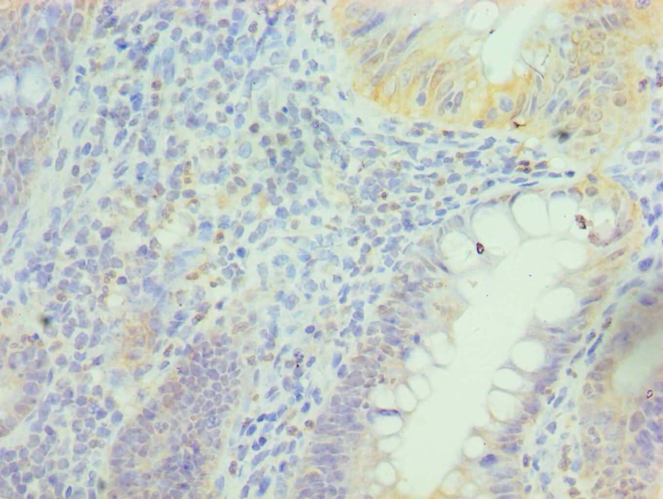
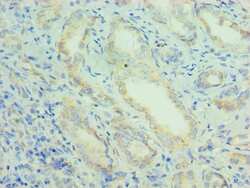
- Immunohistochemical analysis of PRDX5 in paraffin embedded human kidney tissue using a PRDX5 polyclonal antibody (Product # PA5-98085) at a dilution of 1:100.
- Submitted by
- Invitrogen Antibodies (provider)
- Main image

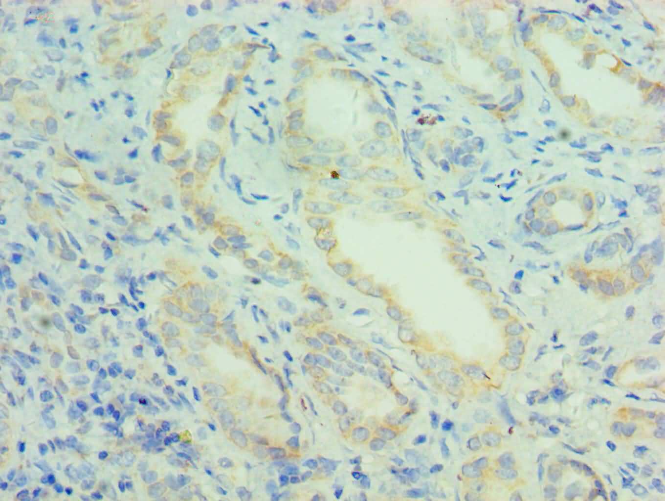
- Experimental details
- Immunohistochemical analysis of PRDX5 in paraffin embedded human kidney tissue using a PRDX5 polyclonal antibody (Product # PA5-98085) at a dilution of 1:100.

Supportive validation
- Submitted by
- Invitrogen Antibodies (provider)
- Main image
- Experimental details
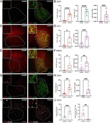
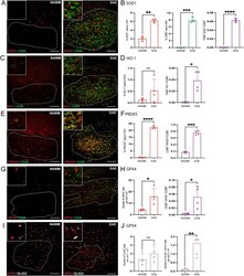
- Expression of antioxidant enzymes is upregulated in POVPC-induced spinal cord lesions. ( A , C , E , G , I ) Representative confocal images of NAWM or POVPC injected spinal cord labeled with CD68 for microglia/macrophage (green), antioxidant enzyme of interest (red), and OLIG2 for oligodendrocyte lineage cells (white). Dotted line indicates the NAWM or lesion region of interest (ROI) selected for image analysis. ( B , D , F ) Bar graphs comparing the percent of ROI that is CD68 + , antioxidant enzyme of interest, and CD68 + antioxidant enzyme + . ( H ) Bar graph comparing the number of GPX4 + cells per mm 2 x 10 2 of ROI and percent of ROI that is CD68 + GPX4 + ( J ) Bar graph representing number of OLIG2 + cells per mm 2 x 10 2 of ROI and percent of ROI that is OLIG2 + GPX4 + . Scale bar = 100 um. Data are shown as mean +- S.D, n = 6-12 mice. Significance indicated as * p < 0.05, ** p < 0.01, *** p < 0.001, **** p < 0.0001, two-tailed, paired student's t-test.
- Submitted by
- Invitrogen Antibodies (provider)
- Main image
- Experimental details
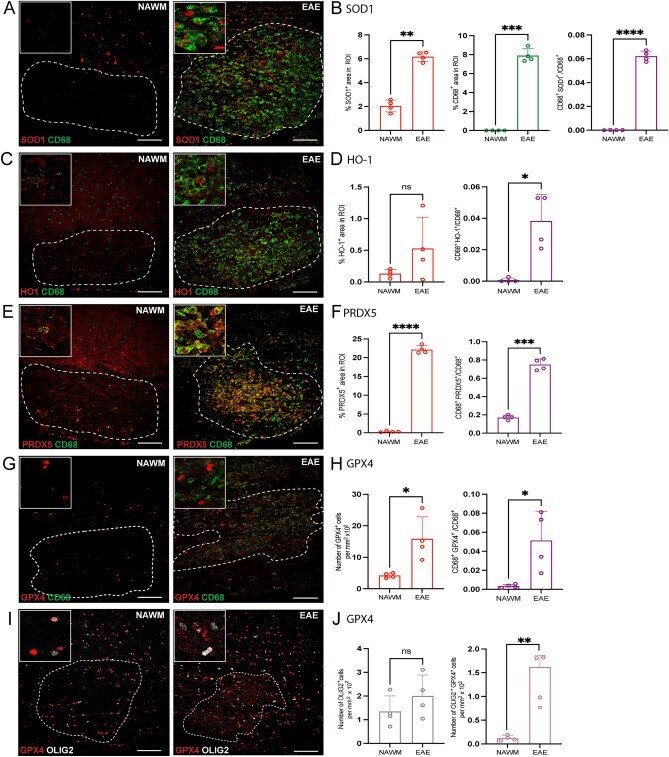
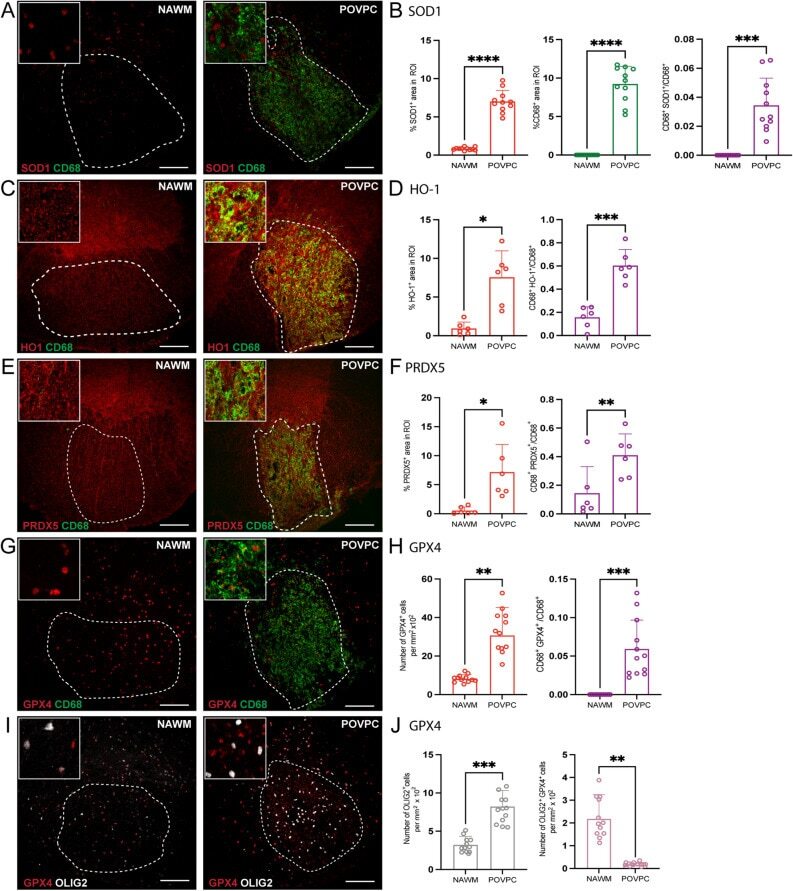
- Detection of antioxidant enzymes is increased in EAE-induced lesions. ( A , C , E , G , I ) Representative confocal images of NAWM or EAE-induced mice labeled with CD68 for microglia/macrophage (green), antioxidant enzyme of interest (red), OLIG2 for oligodendrocyte lineage cells (white). Dotted line indicates the lesion ROI selected for image analysis. ( B , D , F ) Bar graphs comparing the percent of ROI that is CD68 + , antioxidant enzyme of interest, and CD68 + antioxidant enzyme + . ( H ) Bar graph comparing the number of GPX4 + cells per mm 2 x 10 2 of ROI and percent of ROI that is CD68 + GPX4 + ( J ) Bar graph representing number of OLIG2 + cells per mm 2 x 10 2 of ROI and percent of ROI that is OLIG2 + GPX4 + . Scale bar = 100 um. Data are shown as mean +- S.D, n = 4 mice. Significance indicated as * p < 0.05, ** p < 0.01, *** p < 0.001, **** p < 0.0001, two-tailed, paired student's t-test.