Explore
 Validate
 Learn
LearnAF5724
antibody from R&D Systems
Targeting: PRDX5
ACR1, AOEB166, B166, MGC117264, MGC142283, MGC142285, PLP, PMP20, PRDX6, PRXV, SBBI10
 Western blot
Antibody data
- Antibody Data
- Antigen structure
- References [0]
- Comments [0]
- Validations
- Western blot [1]
- Immunocytochemistry [2]
Submit
Validation data
Reference
Comment
Report error
- Product number
- AF5724 - Provider product page

- Provider
- R&D Systems
- Product name
- Human/Mouse/Rat Peroxiredoxin 5 Antibody
- Antibody type
- Polyclonal
- Description
- Antigen Affinity-purified. Detects human, mouse and rat Peroxiredoxin 5 in Western blots.
- Reactivity
- Human, Mouse, Rat
- Host
- Goat
- Conjugate
- Unconjugated
- Antigen sequence
P99029- Isotype
- IgG
- Vial size
- 100 ug
- Concentration
- LYOPH
- Storage
- Use a manual defrost freezer and avoid repeated freeze-thaw cycles. 12 months from date of receipt, -20 to -70 °C as supplied. 1 month, 2 to 8 °C under sterile conditions after reconstitution. 6 months, -20 to -70 °C under sterile conditions after reconstitution.
No comments: Submit comment
Supportive validation
- Submitted by
- R&D Systems (provider)
- Main image
- Experimental details
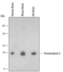
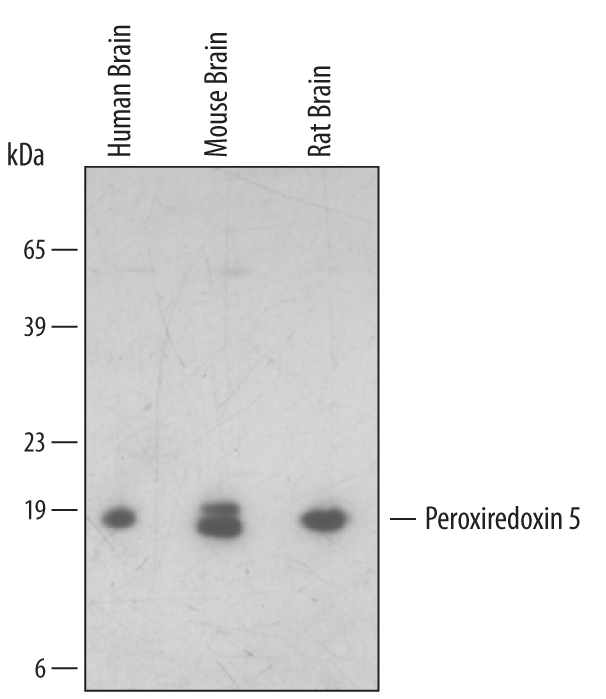
- Detection of Human/Mouse/Rat Peroxiredoxin 5 by Western Blot. Western blot shows lysates of human, mouse, and rat brain tissue. PVDF membrane was probed with 1 µg/mL of Goat Anti-Human/Mouse/Rat Peroxiredoxin 5 Antigen Affinity-purified Polyclonal Antibody (Catalog # AF5724) followed by HRP-conjugated Anti-Goat IgG Secondary Antibody (Catalog # HAF109). A specific band was detected for Peroxiredoxin 5 at approximately 17 kDa (as indicated). This experiment was conducted under reducing conditions and using Immunoblot Buffer Group 2.
Supportive validation
- Submitted by
- R&D Systems (provider)
- Main image

- Experimental details
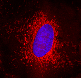
- Peroxiredoxin 5 in A549 Human Cell Line. Peroxiredoxin 5 was detected in immersion fixed A549 human lung carcinoma cell line using Goat Anti-Human/Mouse/Rat Peroxiredoxin 5 Antigen Affinity-purified Polyclonal Antibody (Catalog # AF5724) at 15 µg/mL for 3 hours at room temperature. Cells were stained using the NorthernLights™ 557-conjugated Anti-Goat IgG Secondary Antibody (red; Catalog # NL001) and counterstained with DAPI (blue). Specific staining was localized to cytoplasm (mitochondria). View our protocol for Fluorescent ICC Staining of Cells on Coverslips.
- Submitted by
- R&D Systems (provider)
- Main image

- Experimental details
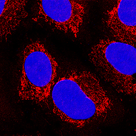
- Peroxiredoxin 5 in A549 Human Cell Line. Peroxiredoxin 5 was detected in immersion fixed A549 human lung carcinoma cell line using Goat Anti-Human/Mouse/Rat Peroxiredoxin 5 Antigen Affinity-purified Polyclonal Antibody (Catalog # AF5724) at 10 µg/mL for 3 hours at room temperature. Cells were stained using the NorthernLights™ 557-conjugated Anti-Goat IgG Secondary Antibody (red; Catalog # NL001) and counterstained with DAPI (blue). Specific staining was localized to mitochondria. View our protocol for Fluorescent ICC Staining of Cells on Coverslips.