Explore
 Validate
 Learn
Learn Western blot
 Immunocytochemistry
Antibody data
- Antibody Data
- Antigen structure
- References [2]
- Comments [0]
- Validations
- Immunocytochemistry [5]
- Immunohistochemistry [4]
- Chromatin Immunoprecipitation [2]
- Other assay [1]
Submit
Validation data
Reference
Comment
Report error
- Product number
- MA5-18108 - Provider product page

- Provider
- Invitrogen Antibodies
- Product name
- EZH2 Monoclonal Antibody (144CT2.1.1.5)
- Antibody type
- Monoclonal
- Antigen
- Recombinant full-length protein
- Description
- This antibody is predicted to react with non-human primate based on sequence homology.
- Reactivity
- Human
- Host
- Mouse
- Isotype
- IgG
- Antibody clone number
- 144CT2.1.1.5
- Vial size
- 400 μL
- Concentration
- 0.48 mg/mL
- Storage
- Store at 4°C short term. For long term storage, store at -20°C, avoiding freeze/thaw cycles.
Submitted references Quantifying the phase separation property of chromatin-associated proteins under physiological conditions using an anti-1,6-hexanediol index.
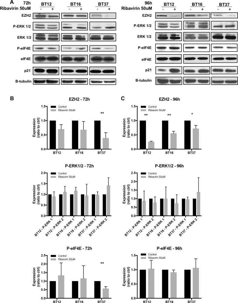
Ribavirin as a potential therapeutic for atypical teratoid/rhabdoid tumors.
Shi M, You K, Chen T, Hou C, Liang Z, Liu M, Wang J, Wei T, Qin J, Chen Y, Zhang MQ, Li T
Genome biology 2021 Aug 17;22(1):229
Genome biology 2021 Aug 17;22(1):229
Ribavirin as a potential therapeutic for atypical teratoid/rhabdoid tumors.
Casaos J, Huq S, Lott T, Felder R, Choi J, Gorelick N, Peters M, Xia Y, Maxwell R, Zhao T, Ji C, Simon T, Sesen J, Scotland SJ, Kast RE, Rubens J, Raabe E, Eberhart CG, Jackson EM, Brem H, Tyler B, Skuli N
Oncotarget 2018 Jan 30;9(8):8054-8067
Oncotarget 2018 Jan 30;9(8):8054-8067
No comments: Submit comment
Supportive validation
- Submitted by
- Invitrogen Antibodies (provider)
- Main image
- Experimental details
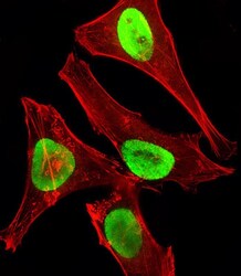
- Immunofluorescent analysis of EZH2 showing staining in the nucleus of U-251 cells using an EZH2 monoclonal antibody (Product # MA5-18108) followed by detection using a fluorescent conjugated secondary antibody (green). Cytoplasmic actin was stained with a fluorescent red phalloidin (7units/mL, 1 h at 37ºC).

- Submitted by
- Invitrogen Antibodies (provider)
- Main image
- Experimental details
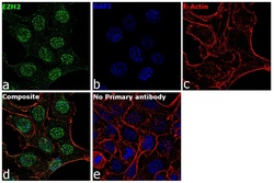
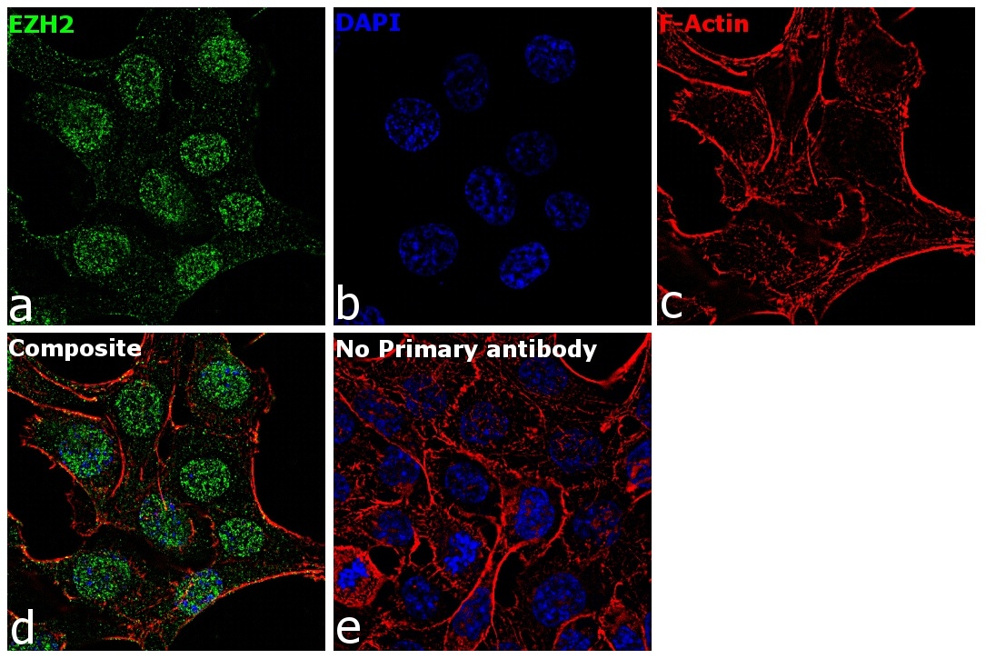
- Immunofluorescence analysis of EZH2 was performed using 70% confluent log phase HCT 116 cells. The cells were fixed with 4% paraformaldehyde for 10 minutes, permeabilized with 0.1% Triton™ X-100 for 15 minutes, and blocked with 1% BSA for 1 hour at room temperature. The cells were labeled with EZH2 Mouse Monoclonal Antibody (144CT2.1.1.5) (Product # MA5-18108) at 1: 50 dilution in 0.1% BSA, incubated at 4 degree Celsius overnight and then labeled with Goat anti-Mouse IgG (H+L) Superclonal™ Secondary Antibody, Alexa Fluor® 488 conjugate (Product # A28175) at a dilution of 1:2000 for 45 minutes at room temperature (Panel a: green). Nuclei (Panel b: blue) were stained with ProLong™ Diamond Antifade Mountant with DAPI (Product # P36962). F-actin (Panel c: red) was stained with Rhodamine Phalloidin (Product # R415, 1:300). Panel d represents the merged image showing nucleus localization. Panel e represents control cells with no primary antibody to assess background. The images were captured at 60X magnification.

- Submitted by
- Invitrogen Antibodies (provider)
- Main image

- Experimental details
- Immunofluorescence analysis of EZH2 was performed using 70% confluent log phase HCT 116 cells. The cells were fixed with 4% paraformaldehyde for 10 minutes, permeabilized with 0.1% Triton™ X-100 for 15 minutes, and blocked with 1% BSA for 1 hour at room temperature. The cells were labeled with EZH2 Mouse Monoclonal Antibody (144CT2.1.1.5) (Product # MA5-18108) at 1: 50 dilution in 0.1% BSA, incubated at 4 degree Celsius overnight and then labeled with Goat anti-Mouse IgG (H+L) Superclonal™ Secondary Antibody, Alexa Fluor® 488 conjugate (Product # A28175) at a dilution of 1:2000 for 45 minutes at room temperature (Panel a: green). Nuclei (Panel b: blue) were stained with ProLong™ Diamond Antifade Mountant with DAPI (Product # P36962). F-actin (Panel c: red) was stained with Rhodamine Phalloidin (Product # R415, 1:300). Panel d represents the merged image showing nucleus localization. Panel e represents control cells with no primary antibody to assess background. The images were captured at 60X magnification.
- Submitted by
- Invitrogen Antibodies (provider)
- Main image

- Experimental details
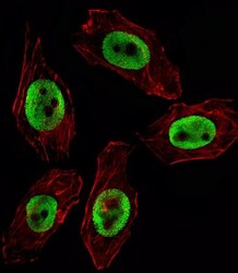
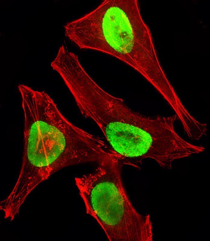
- Immunocytochemistry analysis of EZH2 in U251 cells. Samples were incubated with EZH2 monoclonal antibody (Product # MA5-18108) using a dilution of 1:25 for 1 h at 37°C followed by Alexa Fluor® 488 conjugated donkey anti-mouse antibody (green) at a dilution of 1:400 for 50 min at 37°C. Cells were fixed with 4% PFA (20 min) and permeabilized with Triton X-100 (0.1%, 10 min). Cytoplasmic actin was counterstained with Alexa Fluor® 555 (red) conjugated Phalloidin (7 units/mL, 1 h at 37°C). EZH2 immunoreactivity is localized to Nucleus significantly.

- Submitted by
- Invitrogen Antibodies (provider)
- Main image
- Experimental details
- Immunocytochemistry analysis of EZH2 in Hela (Human Cervical epithelial adenocarcinoma cell line) cells. Samples were incubated with EZH2 monoclonal antibody (Product # MA5-18108) using a dilution of 1:25 followed by Dylight® 488-conjugated goat anti-mouse IgG at a dilution of 1:200 (green). Cells were 4% paraformaldehyde-fixed and 0.1% Triton X-100 permeabilized. Immunofluorescence image showing nucleus staining on HeLa cell line. Cytoplasmic actin is detected with Dylight® 554 Phalloidin at 1:100 dilution (red).
Supportive validation
- Submitted by
- Invitrogen Antibodies (provider)
- Main image
- Experimental details
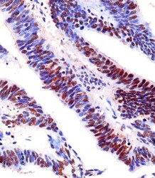
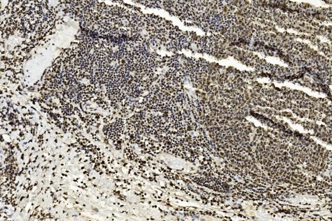
- Immunohistochemistry analysis of EZH2 in paraformaldehyde-fixed, paraffin-embedded human colorectal carcinoma tissue sections. Samples were incubated with EZH2 monoclonal antibody (Product # MA5-18108) using a dilution of 1:25 for 1 hours at 37°C followed by an undiluted biotinylated goat polyvalent antibody. Tissue was fixed with formaldehyde and blocked with 3% BSA for 0.5 hour at room temperature; antigen retrieval was by heat mediation with a citrate buffer (pH 6).

- Submitted by
- Invitrogen Antibodies (provider)
- Main image
- Experimental details
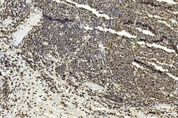
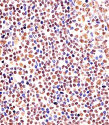
- Immunohistochemistry analysis of EZH2 in paraffin-embedded Human tonsil section. Samples were incubated with EZH2 monoclonal antibody (Product # MA5-18108) using a dilution of 0.0763888888888889 followed by an undiluted biotinylated goat polyvalent antibody was used as the secondary, followed by DAB staining.
- Submitted by
- Invitrogen Antibodies (provider)
- Main image

- Experimental details
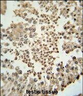
- Immunohistochemistry analysis of EZH2 in formalin fixed and paraffin embedded human testis tissue. Samples were incubated with EZH2 monoclonal antibody (Product # MA5-18108) followed by peroxidase conjugation of the secondary antibody and DAB staining. This data demonstrates the use of this antibody for immunohistochemistry. Clinical relevance has not been evaluated.
- Submitted by
- Invitrogen Antibodies (provider)
- Main image
- Experimental details
- Immunohistochemistry analysis of EZH2 in paraformaldehyde-fixed paraffin embedded human tonsil tissue sections . Samples were incubated with EZH2 monoclonal antibody (Product # MA5-18108) using a dilution of 1:25 for 1 hours at 37°C followed by an undiluted biotinylated goat polyvalent antibody. Tissue was fixed with formaldehyde and blocked with 3% BSA for 0.5 hour at room temperature; antigen retrieval was by heat mediation with a citrate buffer (pH 6).

Supportive validation
- Submitted by
- Invitrogen Antibodies (provider)
- Main image
- Experimental details
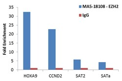
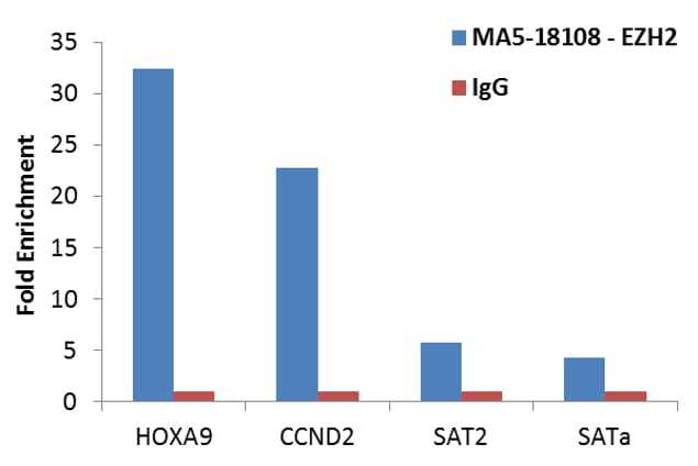
- Enrichment of endogenous EZH2 protein at specific gene loci using Anti-EZH2 Antibody: Chromatin Immunoprecipitation (ChIP) was performed using Anti-EZH2 mouse monoclonal antibody (Product # MA5-18108, 8 ul) on sheared chromatin from 2 million HCT 116 cells using the MAGnify ChIP System (Product # 49-2024). Normal Rabbit IgG was used as a negative IP control. The purified DNA was analyzed by qPCR with PCR primer pairs over the promoters of HOXA9, CCND2 (positive) and SAT2 satellite repeats and SATa satellite alpha (negative). Data is presented as fold enrichment of the antibody signal versus the negative control IgG using the comparative CT method.

- Submitted by
- Invitrogen Antibodies (provider)
- Main image

- Experimental details
- Enrichment of endogenous EZH2 protein at specific gene loci using Anti-EZH2 Antibody: Chromatin Immunoprecipitation (ChIP) was performed using Anti-EZH2 mouse monoclonal antibody (Product # MA5-18108, 8 ul) on sheared chromatin from 2 million HCT 116 cells using the MAGnify ChIP System (Product # 49-2024). Normal Rabbit IgG was used as a negative IP control. The purified DNA was analyzed by qPCR with PCR primer pairs over the promoters of HOXA9, CCND2 (positive) and SAT2 satellite repeats and SATa satellite alpha (negative). Data is presented as fold enrichment of the antibody signal versus the negative control IgG using the comparative CT method.
Supportive validation
- Submitted by
- Invitrogen Antibodies (provider)
- Main image

- Experimental details
- NULL