Explore
 Validate
 Learn
Learn Western blot
Antibody data
- Antibody Data
- Antigen structure
- References [1]
- Comments [0]
- Validations
- Western blot [1]
Submit
Validation data
Reference
Comment
Report error
- Product number
- HPA041939 - Provider product page

- Provider
- Atlas Antibodies
- Proper citation
- Atlas Antibodies Cat#HPA041939, RRID:AB_10793938
- Product name
- Anti-PRPF31
- Antibody type
- Polyclonal
- Description
- Polyclonal Antibody against Human PRPF31, Gene description: pre-mRNA processing factor 31, Alternative Gene Names: hPrp31, NY-BR-99, PRP31, RP11, SNRNP61, Validated applications: WB, Uniprot ID: Q8WWY3, Storage: Store at +4°C for short term storage. Long time storage is recommended at -20°C.
- Reactivity
- Human
- Host
- Rabbit
- Conjugate
- Unconjugated
- Isotype
- IgG
- Vial size
- 100 µl
- Concentration
- 0.1 mg/ml
- Storage
- Store at +4°C for short term storage. Long time storage is recommended at -20°C.
- Handling
- The antibody solution should be gently mixed before use.
Submitted references A Precision Therapy Approach for Retinitis Pigmentosa 11 Using Splice-Switching Antisense Oligonucleotides to Restore the Open Reading Frame of PRPF31
Grainok J, Pitout I, Chen F, McLenachan S, Heath Jeffery R, Mitrpant C, Fletcher S
International Journal of Molecular Sciences 2024;25(6):3391
International Journal of Molecular Sciences 2024;25(6):3391
No comments: Submit comment
Enhanced validation
- Submitted by
- klas2
- Enhanced method
- Genetic validation
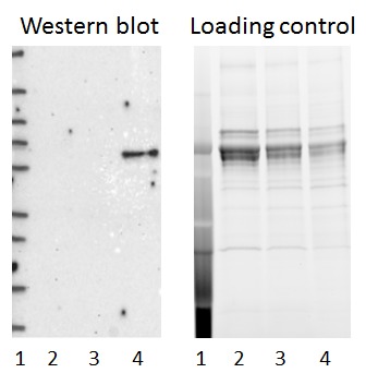
- Main image
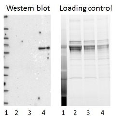
- Experimental details
- Western blot of cell lysate from U-2 OS cells transfected with either siRNA targeting PRPF31 or control siRNA. Lane 1: Marker (250, 130, 95, 72, 55, 36, 28, 17, 10) Lane 2: Cell lysate from U-2OS cells transfected with siRNA targeting PRPF31 Lane 3: N/A Lane 4: Cell lysate from U-2OS cells transfected with control siRNA Right image, lane 1-4: loading control
- Sample type
- U-2 OS
- Primary Ab dilution
- 1:239
- Conjugate
- Horseradish Peroxidase
- Secondary Ab
- Secondary Ab
- Secondary Ab dilution
- 1:3000
- Knockdown/Genetic Approaches Application
- Western blot