 Explore
 Validate
 Learn
Learn Immunocytochemistry
Antibody data
- Antibody Data
- Antigen structure
- References [1]
- Comments [0]
- Validations
- Immunocytochemistry [4]
- Immunohistochemistry [1]
- Flow cytometry [1]
Submit
Validation data
Reference
Comment
Report error
- Product number
- 700242 - Provider product page

- Provider
- Invitrogen Antibodies
- Product name
- Phospho-MNK1 (Thr197, Thr202) Recombinant Rabbit Monoclonal Antibody (18H4L11)
- Antibody type
- Monoclonal
- Antigen
- Synthetic peptide
- Description
- This antibody is predicted to react with bovine, canine, chicken, chimpanzee, equine, mouse, primate, rat, Rhesus monkey , porcine, Xenopus and zebrafish based on sequence homology. Intact IgG appears on a non-reducing gel as ~150 kDa band and upon reduction generating a ~25 kDa light chain band and a ~50 kDa heavy chain. Recombinant rabbit monoclonal antibodies are produced using in vitro expression systems. The expression systems are developed by cloning in the specific antibody DNA sequences from immunoreactive rabbits. Then, individual clones are screened to select the best candidates for production. The advantages of using recombinant rabbit monoclonal antibodies include: better specificity and sensitivity, lot-to-lot consistency, animal origin-free formulations, and broader immunoreactivity to diverse targets due to larger rabbit immune repertoire.
- Reactivity
- Human
- Host
- Rabbit
- Isotype
- IgG
- Antibody clone number
- 18H4L11
- Vial size
- 100 μg
- Concentration
- 0.5 mg/mL
- Storage
- Store at 4°C short term. For long term storage, store at -20°C, avoiding freeze/thaw cycles.
Submitted references Enteral delivery of proteins stimulates protein synthesis in human duodenal mucosa in the fed state through a mammalian target of rapamycin-independent pathway.
Coëffier M, Claeyssens S, Bôle-Feysot C, Guérin C, Maurer B, Lecleire S, Lavoinne A, Donnadieu N, Cailleux AF, Déchelotte P
The American journal of clinical nutrition 2013 Feb;97(2):286-94
The American journal of clinical nutrition 2013 Feb;97(2):286-94
No comments: Submit comment
Supportive validation
- Submitted by
- Invitrogen Antibodies (provider)
- Main image
- Experimental details
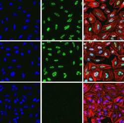
- Immunofluorescent analysis of Phospho-MNK pThr197/202 in HeLa cells using a Phospho-MNK pThr197/202 recombinant rabbit monoclonal antibody (Product # 700242) at a dilution of 5 µg/mL in the absence of peptide (top left) and presence of phosphopeptide used as immunogen (top right) or non-phosphopeptide (bottom left), followed by detection using an Alexa Fluor 488-conjugated goat anti-rabbit secondary antibody at a dilution of 1:1000. Actin was stained with Alexa Fluor 568 phalloidin (Product # A12380). Hoechst only (blue, left), AF488 signal only (green, center) and composite image with Phalloidin (right).

- Submitted by
- Invitrogen Antibodies (provider)
- Main image

- Experimental details
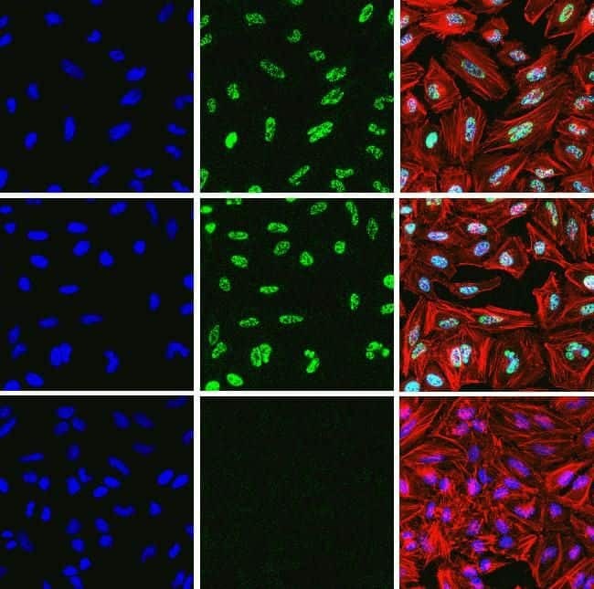
- Immunofluorescent analysis of Phospho-MNK pThr197/202 in HeLa cells using a Phospho-MNK pThr197/202 recombinant rabbit monoclonal antibody (Product # 700242) at a dilution of 5 µg/mL in the absence of peptide (top left) and presence of phosphopeptide used as immunogen (top right) or non-phosphopeptide (bottom left), followed by detection using an Alexa Fluor 488-conjugated goat anti-rabbit secondary antibody at a dilution of 1:1000. Actin was stained with Alexa Fluor 568 phalloidin (Product # A12380). Hoechst only (blue, left), AF488 signal only (green, center) and composite image with Phalloidin (right).
- Submitted by
- Invitrogen Antibodies (provider)
- Main image
- Experimental details
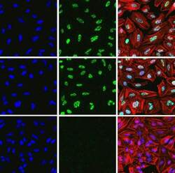
- Immunofluorescent analysis of Phospho-MNK pThr197/202 in HeLa cells using a Phospho-MNK pThr197/202 recombinant rabbit monoclonal antibody (Product # 700242) at a dilution of 5 µg/mL in the absence of peptide (top left) and presence of phosphopeptide used as immunogen (top right) or non-phosphopeptide (bottom left), followed by detection using an Alexa Fluor 488-conjugated goat anti-rabbit secondary antibody at a dilution of 1:1000. Actin was stained with Alexa Fluor 568 phalloidin (Product # A12380). Hoechst only (blue, left), AF488 signal only (green, center) and composite image with Phalloidin (right).
- Submitted by
- Invitrogen Antibodies (provider)
- Main image

- Experimental details
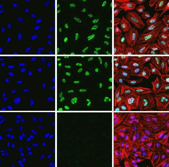
- Immunofluorescent analysis of Phospho-MNK pThr197/202 in HeLa cells using a Phospho-MNK pThr197/202 recombinant rabbit monoclonal antibody (Product # 700242) at a dilution of 5 µg/mL in the absence of peptide (top left) and presence of phosphopeptide used as immunogen (top right) or non-phosphopeptide (bottom left), followed by detection using an Alexa Fluor 488-conjugated goat anti-rabbit secondary antibody at a dilution of 1:1000. Actin was stained with Alexa Fluor 568 phalloidin (Product # A12380). Hoechst only (blue, left), AF488 signal only (green, center) and composite image with Phalloidin (right).

Supportive validation
- Submitted by
- Invitrogen Antibodies (provider)
- Main image
- Experimental details
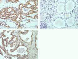
- Immunohistochemistry analysis of Phospho-MNK pThr197/202 in formalin-fixed, paraffin-embedded human thyroid carcinoma (top left), normal thyroid (top right) and prostate carcinoma (bottom) using a Phospho-MNK pThr197/202 monoclonal antibody (Product # 700242) at a dilution of 2 µg/mL. Staining was visualized using DAB and images were taken at a magnification of 20x. Results show nuclear and cytoplasmic staining in tumor cells and no staining in normal tissue.

Supportive validation
- Submitted by
- Invitrogen Antibodies (provider)
- Main image
- Experimental details
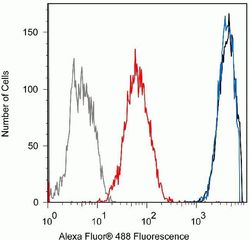
- Flow cytometry analysis of Phospho-MNK pThr197/202 in Jurkat cells using a Phospho-MNK pThr197/202 recombinant rabbit monoclonal antibody (Product # 700242) at a dilution of 0.5ug. Cells were fixed and permeabilized using FIX & PERM (Product # GAS004) reagent, and detection was performed using an Alexa Fluor 488 goat anti-rabbit IgG (black) compared to a control without primary antibody (gray). Pre-incubation with the phosphopeptide decreased the signal (red) whereas incubation with the non-phosphopeptide did not (blue).
