 Explore
 Validate
 Learn
Learn Western blot
 Immunocytochemistry
Antibody data
- Antibody Data
- Antigen structure
- References [0]
- Comments [0]
- Validations
- Western blot [1]
- Immunohistochemistry [1]
- Flow cytometry [2]
Submit
Validation data
Reference
Comment
Report error
- Product number
- NBP2-52983 - Provider product page

- Provider
- Novus Biologicals
- Product name
- Rabbit Polyclonal Thioredoxin-1 Antibody
- Antibody type
- Polyclonal
- Description
- Immunogen affinity purified.
- Reactivity
- Human, Mouse
- Host
- Rabbit
- Isotype
- IgG
- Vial size
- 0.1 ml
- Concentration
- 1.0 mg/ml
- Storage
- Store at 4C short term. Aliquot and store at -20C long term. Avoid freeze-thaw cycles.
No comments: Submit comment
Supportive validation
- Submitted by
- Novus Biologicals (provider)
- Main image

- Experimental details
- Western Blot: Thioredoxin Antibody [NBP2-52983] - Total protein from human HeLa cells, mouse 3T3 cells and rat PC12 cells was separated on a 4-15% gel by SDS-PAGE, transferred to 0.2 um pore size PVDF membrane and blocked in 5% non-fat milk in TBST. The membrane was probed with 2.0 ug/ml anti-Thioredoxin in 1% non-fat milk in TBST and detected with an anti-rabbit HRP secondary antibody using chemiluminescence.

Supportive validation
- Submitted by
- Novus Biologicals (provider)
- Main image
- Experimental details
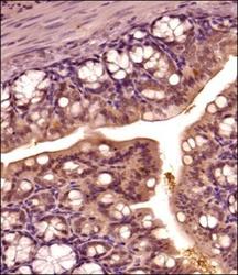
- Immunohistochemistry-Paraffin: Thioredoxin Antibody [NBP2-52983] - IHC analysis of a formalin fixed and paraffin embedded tissue section of mouse intestine using rabbit polyclonal antibody against Thioredoxin (1:300 dilution). The signal was developed with HRP-secondary -DAB method and the sections were counter-stained using hematoxylin. The staining was found primarily in the epithelial cells of the intestinal lumen with weak to negligible signal in the basal or muscular layers. Isolated epithelial cells depicted nuclear positivity also.

Supportive validation
- Submitted by
- Novus Biologicals (provider)
- Main image

- Experimental details
- Flow Cytometry: Thioredoxin-1 Antibody [NBP2-52983] - Thioredoxin Antibody [NBP2-52983] - An intracellular stain was performed on U-87 MG cells with Thioredoxin Antibody NBP2-52983 and a matched isotype control. Cells were fixed with 4% PFA and then permeablized with 0.1% saponin. Cells were incubated in an antibody dilution of 2.5 ug/mL for 30 minutes at room temperature, followed by Rabbit IgG (H+L) Cross-Adsorbed Secondary Antibody.

- Submitted by
- Novus Biologicals (provider)
- Main image

- Experimental details
- Flow Cytometry: Thioredoxin-1 Antibody [NBP2-52983] - An intracellular stain was performed on HeLa cells with Thioredoxin-1 Antibody NBP2-52983AF488 (blue) and a matched isotype control (orange). Cells were fixed with 4% PFA and then permeabilized with 0.1% saponin. Cells were incubated in an antibody dilution of 5 ug/mL for 30 minutes at room temperature. Both antibodies were conjugated to Alexa Fluor 488.
