Explore
 Validate
 Learn
Learn Western blot
Antibody data
- Antibody Data
- Antigen structure
- References [0]
- Comments [0]
- Validations
- Western blot [1]
- Immunocytochemistry [3]
- Immunohistochemistry [3]
- Flow cytometry [1]
Submit
Validation data
Reference
Comment
Report error
- Product number
- PA5-86029 - Provider product page

- Provider
- Invitrogen Antibodies
- Product name
- Thioredoxin 1 Polyclonal Antibody
- Antibody type
- Polyclonal
- Antigen
- Synthetic peptide
- Reactivity
- Human, Mouse
- Host
- Rabbit
- Isotype
- IgG
- Vial size
- 100 µL
- Concentration
- 1 mg/mL
- Storage
- Store at 4°C short term. For long term storage, store at -20°C, avoiding freeze/thaw cycles.
No comments: Submit comment
Supportive validation
- Submitted by
- Invitrogen Antibodies (provider)
- Main image
- Experimental details
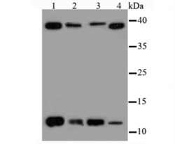
- Western blot analysis of Thioredoxin 1 in different lysates using a Polyclonal antibody (Product #PA5-86029) at a dilution of 1:500. Positive control: Lane1: Hela, Lane 2: HepG2, Lane 3: A431, Lane 4: PC-3M.

Supportive validation
- Submitted by
- Invitrogen Antibodies (provider)
- Main image
- Experimental details
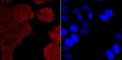
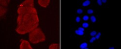
- Immunocytochemical analysis of Thioredoxin 1 in Hela cells using a Thioredoxin 1 Polyclonal antibody (Product # PA5-86029) as seen in red. The nuclear counter stain is DAPI (blue). Cells were fixed in paraformaldehyde, permeabilised with 0.25% Triton X100/PBS.
- Submitted by
- Invitrogen Antibodies (provider)
- Main image
- Experimental details
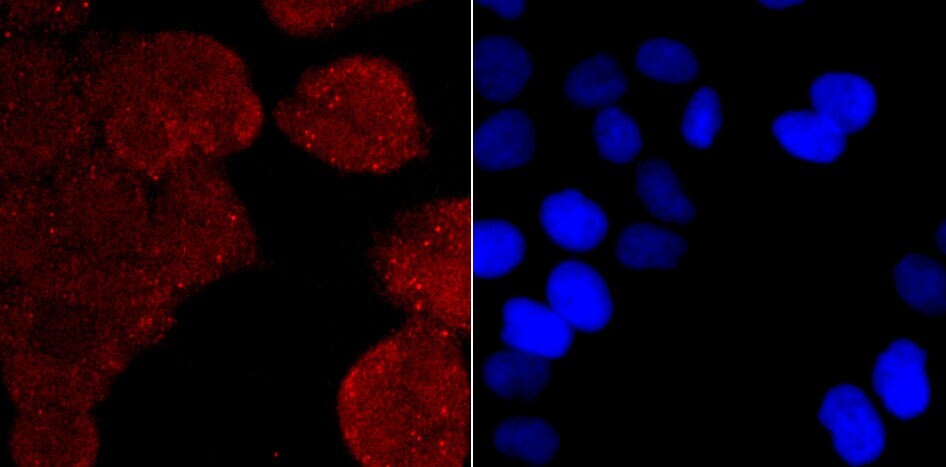
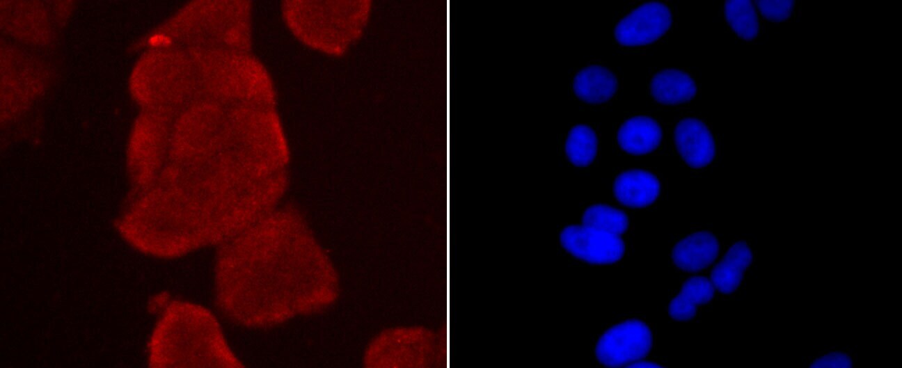
- Immunocytochemical analysis of Thioredoxin 1 in MCF-7 cells using a Thioredoxin 1 Polyclonal antibody (Product # PA5-86029) as seen in red. The nuclear counter stain is DAPI (blue). Cells were fixed in paraformaldehyde, permeabilised with 0.25% Triton X100/PBS.
- Submitted by
- Invitrogen Antibodies (provider)
- Main image

- Experimental details
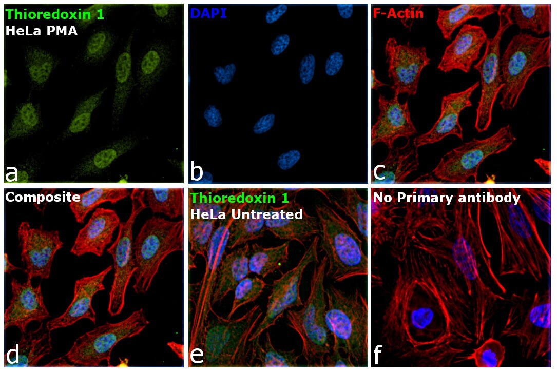
- Immunofluorescence analysis of TXN was performed using 70% confluent log phase HeLa cells treated with PMA (30 ng/mL for 24hrs). The cells were fixed with 4% paraformaldehyde for 10 minutes, permeabilized with 0.1% Triton™ X-100 for 15 minutes, and blocked with 2% BSA for 45 minutes at room temperature. The cells were labeled with Thioredoxin 1 Polyclonal Antibody (Product # PA5-86029) at 1:100 dilution in 0.1% BSA, incubated at 4 degree celsius overnight and then labeled with Donkey anti-Rabbit IgG (H+L) Highly Cross-Adsorbed Secondary Antibody, Alexa Fluor Plus 488 (Product # A32790), (1:2000 dilution), for 45 minutes at room temperature (Panel a: Green). Nuclei (Panel b:Blue) were stained with ProLong™ Diamond Antifade Mountant with DAPI (Product # P36962). F-actin (Panel c: Green) was stained with Rhodamine Phalloidin (Product # R415, 1:300). Panel d represents the merged image showing nuclear localization on PMA treatment and cytoplasmic localization in untreated HeLa cells (Panel e). Panel f represents control cells with no primary antibody to assess the background. The images were captured at 60X magnification.
Supportive validation
- Submitted by
- Invitrogen Antibodies (provider)
- Main image
- Experimental details
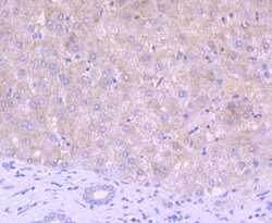
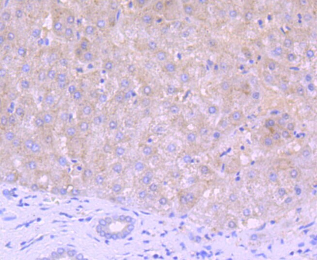
- Immunohistochemical analysis of Thioredoxin 1 of paraffin-embedded Human liver tissue using a Thioredoxin-1 Polyclonal antibody (Product #PA5-86029). Counter stained with hematoxylin.
- Submitted by
- Invitrogen Antibodies (provider)
- Main image

- Experimental details
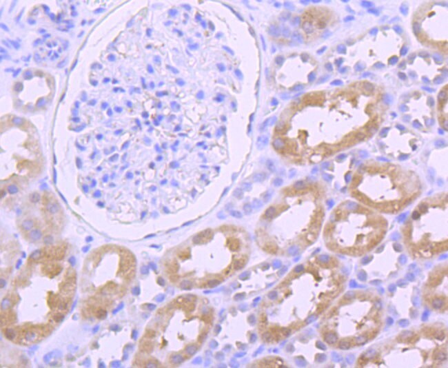
- Immunohistochemical analysis of Thioredoxin 1 of paraffin-embedded Human kidney tissue using a Thioredoxin-1 Polyclonal antibody (Product #PA5-86029). Counter stained with hematoxylin.
- Submitted by
- Invitrogen Antibodies (provider)
- Main image
- Experimental details
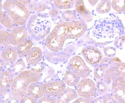
- Immunohistochemical analysis of Thioredoxin 1 of paraffin-embedded Mouse kidney tissue using a Thioredoxin-1 Polyclonal antibody (Product #PA5-86029). Counter stained with hematoxylin.

Supportive validation
- Submitted by
- Invitrogen Antibodies (provider)
- Main image
- Experimental details
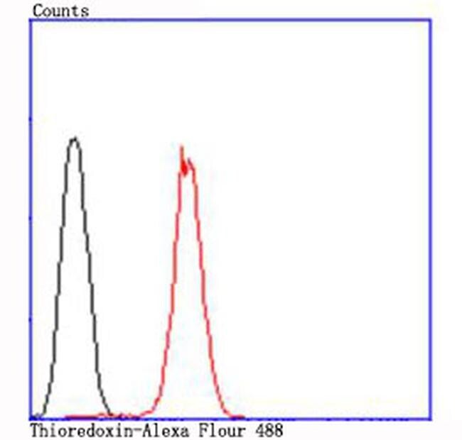
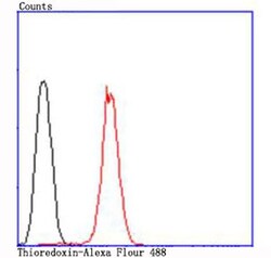
- Flow Cytometric analysis of Thioredoxin 1 in Hela cells using a Thioredoxin 1 Polyclonal Antibody (Product # PA5-86029) at a dilution of 1:100, as seen in red compared with an unlabelled control (cells without incubation with primary antibody; black).