Explore
 Validate
 Learn
Learn Western blot
 Immunocytochemistry
Antibody data
- Antibody Data
- Antigen structure
- References [5]
- Comments [0]
- Validations
- Western blot [5]
- Immunohistochemistry [1]
Submit
Validation data
Reference
Comment
Report error
- Product number
- NB600-533 - Provider product page

- Provider
- Novus Biologicals
- Proper citation
- Novus Cat#NB600-533, RRID:AB_10003448
- Product name
- Rabbit Polyclonal Actin Gamma 1 Antibody
- Antibody type
- Polyclonal
- Description
- Immunogen affinity purified.
- Reactivity
- Human, Mouse
- Host
- Rabbit
- Isotype
- IgG
- Vial size
- 0.1 mg
- Concentration
- 1.0 mg/ml
- Storage
- Store at 4C. Do not freeze.
Submitted references c-Myb and C/EBPβ regulate OPN and other senescence-associated secretory phenotype factors.
Liquid chromatography-mass spectrometry-based quantitative proteomics analysis reveals chondroprotective effects of astragaloside IV in interleukin-1β-induced SW1353 chondrocyte-like cells.
Dual leucine zipper kinase regulates expression of axon guidance genes in mouse neuronal cells.
Okazaki fragment processing-independent role for human Dna2 enzyme during DNA replication.
Human Dna2 is a nuclear and mitochondrial DNA maintenance protein.
Flanagan KC, Alspach E, Pazolli E, Parajuli S, Ren Q, Arthur LL, Tapia R, Stewart SA
Oncotarget 2018 Jan 2;9(1):21-36
Oncotarget 2018 Jan 2;9(1):21-36
Liquid chromatography-mass spectrometry-based quantitative proteomics analysis reveals chondroprotective effects of astragaloside IV in interleukin-1β-induced SW1353 chondrocyte-like cells.
Luo H, Yao L, Zhang Y, Li R
Biomedicine & pharmacotherapy = Biomedecine & pharmacotherapie 2017 Jul;91:796-802
Biomedicine & pharmacotherapy = Biomedecine & pharmacotherapie 2017 Jul;91:796-802
Dual leucine zipper kinase regulates expression of axon guidance genes in mouse neuronal cells.
Blondeau A, Lucier JF, Matteau D, Dumont L, Rodrigue S, Jacques PÉ, Blouin R
Neural development 2016 Jul 28;11(1):13
Neural development 2016 Jul 28;11(1):13
Okazaki fragment processing-independent role for human Dna2 enzyme during DNA replication.
Duxin JP, Moore HR, Sidorova J, Karanja K, Honaker Y, Dao B, Piwnica-Worms H, Campbell JL, Monnat RJ Jr, Stewart SA
The Journal of biological chemistry 2012 Jun 22;287(26):21980-91
The Journal of biological chemistry 2012 Jun 22;287(26):21980-91
Human Dna2 is a nuclear and mitochondrial DNA maintenance protein.
Duxin JP, Dao B, Martinsson P, Rajala N, Guittat L, Campbell JL, Spelbrink JN, Stewart SA
Molecular and cellular biology 2009 Aug;29(15):4274-82
Molecular and cellular biology 2009 Aug;29(15):4274-82
No comments: Submit comment
Supportive validation
- Submitted by
- Novus Biologicals (provider)
- Main image
- Experimental details
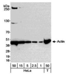
- Western Blot: Actin Gamma 1 Antibody [NB600-533] - Whole cell lysate from HeLa (1, 2.5, 5, 15 and 50 ug) and mouse NIH3T3 cells (50 ug), probed with diluted at 0.04 ug/ml.
- Submitted by
- Novus Biologicals (provider)
- Main image

- Experimental details
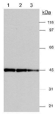
- Western Blot: Actin Gamma 1 Antibody [NB600-533] - Actin Antibody [NB600-533]-Detection of actin in 3T3 (20 ug) lysates. ECL detection 30 seconds. Lane 1 - 1:5,000 dilution Lane 2 - 1:10,000 dilution Lane 3 - 1:15,000 dilution
- Submitted by
- Novus Biologicals (provider)
- Main image

- Experimental details
- Simple Western: Actin Gamma 1 Antibody [NB600-533] - Simple Western lane view shows a specific band for Actin Gamma 1 in 1.0 mg/ml of HeLa lysate. This experiment was performed under reducing conditions using the 12-230kDa separation system.

- Submitted by
- Novus Biologicals (provider)
- Main image
- Experimental details
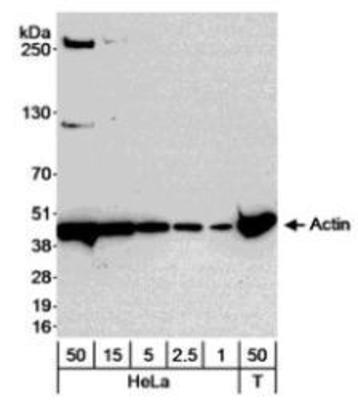
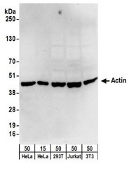
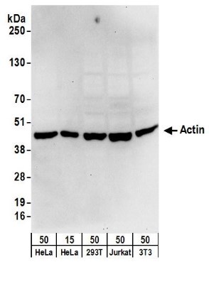
- Western Blot: Actin Gamma 1 Antibody [NB600-533] - Samples: Whole cell lysate from HeLa (15 and 50 ug), 293T (50ug), Jurkat (50ug), and mouse NIH3T3 (50ug) cells. Antibodies: Affinity purified rabbit anti-Actin antibody used for WB at 0.1 ug/ml. Detection: Chemiluminescence with an exposure time of 30 seconds.
- Submitted by
- Novus Biologicals (provider)
- Main image
- Experimental details
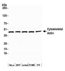
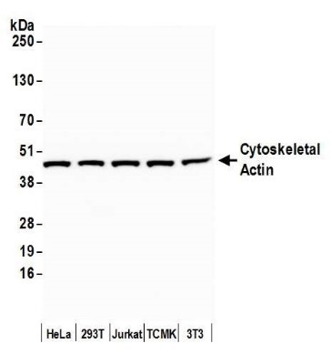
- Western Blot: Actin Gamma 1 Antibody [NB600-533] - Detection of human and mouse Cytoskeletal Actin by western blot. Samples: Whole cell lysate (15 ug) from HeLa, HEK293T, Jurkat, mouse TCMK-1, and mouse NIH 3T3 cells prepared using NETN lysis buffer. Antibody: Affinity purified rabbit anti-Cytoskeletal Actin antibody NB600-533 used for WB at 0.1 ug/ml. Detection: Chemiluminescence with an exposure time of 10 seconds.
Supportive validation
- Submitted by
- Novus Biologicals (provider)
- Main image
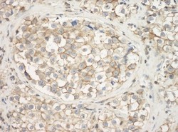
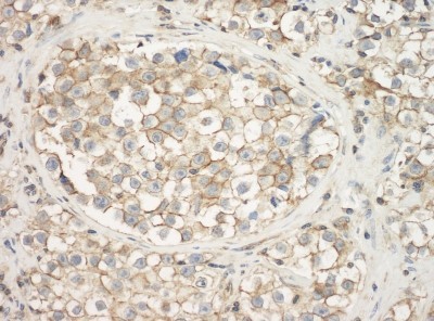
- Experimental details
- Immunohistochemistry: Actin Gamma 1 Antibody [NB600-533] - Sample: FFPE section of human testicular seminoma. Antibody: Affinity purified rabbit anti-Actin used at a dilution of 1:1,000 (1ug/ml). Detection: DAB