Explore
 Validate
 Learn
LearnLF-MA0144
antibody from Invitrogen Antibodies
Targeting: PRDX2
MGC4104, NKEFB, PRP, PRX2, PRXII, TDPX1, TSA
 Western blot
 ELISA
ELISA Immunocytochemistry
Antibody data
- Antibody Data
- Antigen structure
- References [5]
- Comments [0]
- Validations
- Western blot [1]
- Flow cytometry [1]
Submit
Validation data
Reference
Comment
Report error
- Product number
- LF-MA0144 - Provider product page

- Provider
- Invitrogen Antibodies
- Product name
- Anti-PRDX2 Monoclonal Antibody (1E8)
- Antibody type
- Monoclonal
- Antigen
- Recombinant full-length protein
- Description
- A suggested positive control for this product is HeLa cells.
- Reactivity
- Human, Mouse, Rat
- Host
- Mouse
- Isotype
- IgG
- Antibody clone number
- 1E8
- Vial size
- 100 µL
- Storage
- -20° C, Avoid Freeze/Thaw Cycles
Submitted references PRDX2 protects against oxidative stress induced by H. pylori and promotes resistance to cisplatin in gastric cancer.
Highly Purified Human Extracellular Vesicles Produced by Stem Cells Alleviate Aging Cellular Phenotypes of Senescent Human Cells.
Peroxidase expression is decreased by palmitate in cultured podocytes but increased in podocytes of advanced diabetic nephropathy.
Insulin-stimulated lipid accumulation is inhibited by ROS-scavenging chemicals, but not by the Drp1 inhibitor Mdivi-1.
Aldose reductase deficiency leads to oxidative stress-induced dopaminergic neuronal loss and autophagic abnormality in an animal model of Parkinson's disease.
Wang S, Chen Z, Zhu S, Lu H, Peng D, Soutto M, Naz H, Peek R Jr, Xu H, Zaika A, Xu Z, El-Rifai W
Redox biology 2020 Jan;28:101319
Redox biology 2020 Jan;28:101319
Highly Purified Human Extracellular Vesicles Produced by Stem Cells Alleviate Aging Cellular Phenotypes of Senescent Human Cells.
Liu S, Mahairaki V, Bai H, Ding Z, Li J, Witwer KW, Cheng L
Stem cells (Dayton, Ohio) 2019 Jun;37(6):779-790
Stem cells (Dayton, Ohio) 2019 Jun;37(6):779-790
Peroxidase expression is decreased by palmitate in cultured podocytes but increased in podocytes of advanced diabetic nephropathy.
Lee E, Lee HS
Journal of cellular physiology 2018 Dec;233(12):9060-9069
Journal of cellular physiology 2018 Dec;233(12):9060-9069
Insulin-stimulated lipid accumulation is inhibited by ROS-scavenging chemicals, but not by the Drp1 inhibitor Mdivi-1.
Kim JH, Park SJ, Kim B, Choe YG, Lee DS
PloS one 2017;12(10):e0185764
PloS one 2017;12(10):e0185764
Aldose reductase deficiency leads to oxidative stress-induced dopaminergic neuronal loss and autophagic abnormality in an animal model of Parkinson's disease.
Yeung PKK, Lai AKW, Son HJ, Zhang X, Hwang O, Chung SSM, Chung SK
Neurobiology of aging 2017 Feb;50:119-133
Neurobiology of aging 2017 Feb;50:119-133
No comments: Submit comment
Supportive validation
- Submitted by
- Invitrogen Antibodies (provider)
- Main image

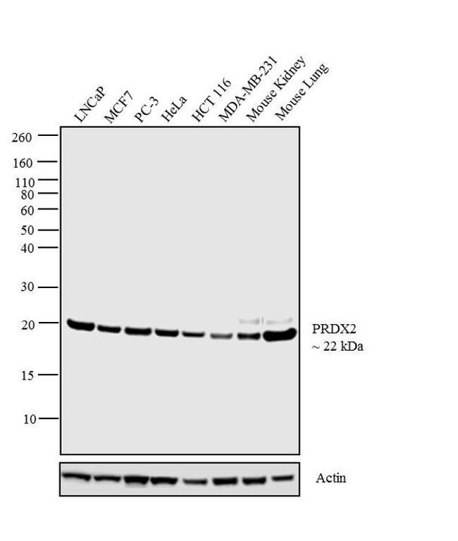
- Experimental details
- Western blot analysis was performed on membrane enriched extracts (30 µg lysate) of LNCaP (Lane 1), MCF7 (Lane 2), PC-3 (Lane 3), HeLa (Lane 4), HCT 116 (Lane 5), MDA-MB-231 (Lane 6), tissue extracts (30 µg lysate) of Mouse Kidney (Lane 7) and Mouse Lung (Lane 8). The blot was probed with Anti-PRDX2 Mouse Monoclonal Antibody (Product # LF-MA0144, 1:3,000 dilution) and detected by chemiluminescence using Goat anti-Mouse IgG (H+L) Superclonal™ Secondary Antibody, HRP conjugate (Product # A28177, 0.4 µg/mL, 1:2500 dilution). A 22 kDa band corresponding to PRDX2 was observed across the cell lines and tissues tested. Known quantity of protein samples were electrophoresed using Novex® NuPAGE® 4-12 % Bis-Tris gel (Product # NP0321BOX), XCell SureLock™ Electrophoresis System (Product # EI0002) and Novex® Sharp Pre-Stained Protein Standard (Product # LC5800). Resolved proteins were then transferred onto a nitrocellulose membrane with iBlot® 2 Dry Blotting System (Product # IB21001). The membrane was probed with the relevant primary and secondary Antibody following blocking with 5 % skimmed milk. Chemiluminescent detection was performed using Pierce™ ECL Western Blotting Substrate (Product # 32106).
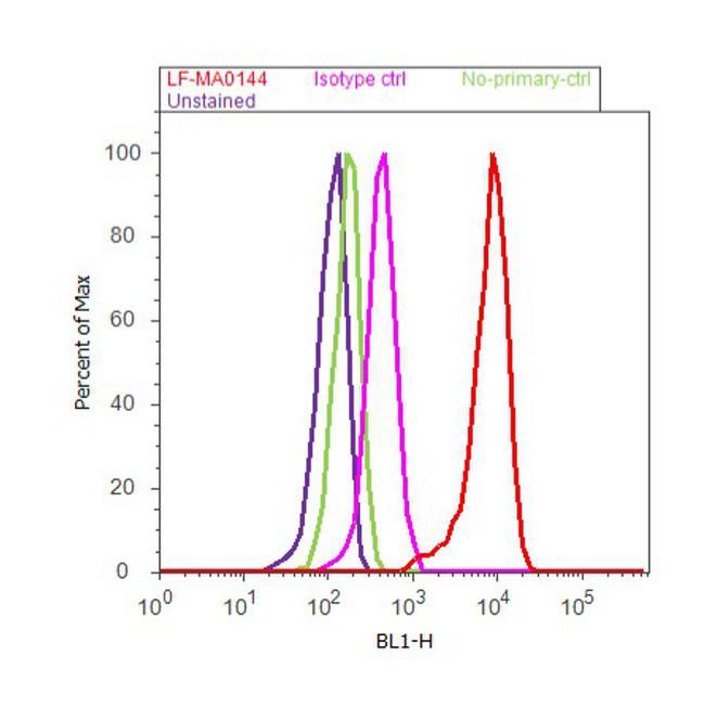
Supportive validation
- Submitted by
- Invitrogen Antibodies (provider)
- Main image

- Experimental details
- Flow cytometry analysis of Peroxiredoxin 4 was done on LNCap cells. Cells were fixed with 70% ethanol for 10 minutes, permeabilized with 0.25% Triton™ X-100 for 20 minutes, and blocked with 5% BSA for 30 minutes at room temperature. Cells were labeled with Peroxiredoxin 4 Mouse Monoclonal Antibody (Product # LF-MA0014, red histogram) at a dilution of 1:20 or a matched isotype control (pink histogram) in 2.5% BSA. After incubation at room temperature for 2 hours, the cells were labeled with Alexa Fluor® 488 Rabbit Anti-Mouse Secondary Antibody (Product # A-11059) at a dilution of 1:400 for 30 minutes at room temperature. The representative 10,000 cells were acquired and analyzed for each sample using an Attune® Acoustic Focusing Cytometer. The purple histogram represents unstained control cells and the green histogram represents no-primary-antibody control.