Explore
 Validate
 Learn
Learn Western blot
Antibody data
- Antibody Data
- Antigen structure
- References [1]
- Comments [0]
- Validations
- Western blot [2]
Submit
Validation data
Reference
Comment
Report error
- Product number
- AF3489 - Provider product page

- Provider
- R&D Systems
- Product name
- Human/Mouse/Rat Peroxiredoxin 2 Antibody
- Antibody type
- Polyclonal
- Description
- Antigen Affinity-purified. The antibody detects endogenous human, mouse and rat Peroxiredoxin 2 in in Western blots. In Western blots, approximately 25% cross-reactivity with recombinant human (rh) Peroxiredoxin 1 and rhPeroxiredoxin 4 is observed, approximately 10% cross-reactivity with rhPeroxiredoxin 3 is observed, and less than 1% cross-reactivity with rhPeroxiredoxin 5 and rhPeroxiredoxin 6 is observed.
- Reactivity
- Human, Mouse, Rat
- Host
- Goat
- Conjugate
- Unconjugated
- Antigen sequence
P32119- Isotype
- IgG
- Vial size
- 100 ug
- Concentration
- LYOPH
- Storage
- Use a manual defrost freezer and avoid repeated freeze-thaw cycles. 12 months from date of receipt, -20 to -70 °C as supplied. 1 month, 2 to 8 °C under sterile conditions after reconstitution. 6 months, -20 to -70 °C under sterile conditions after reconstitution.
Submitted references Peroxiredoxin-2 and STAT3 form a redox relay for H2O2 signaling.
Sobotta MC, Liou W, Stöcker S, Talwar D, Oehler M, Ruppert T, Scharf AN, Dick TP
Nature chemical biology 2015 Jan;11(1):64-70
Nature chemical biology 2015 Jan;11(1):64-70
No comments: Submit comment
Supportive validation
- Submitted by
- R&D Systems (provider)
- Main image
- Experimental details
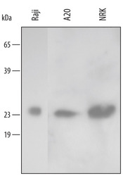
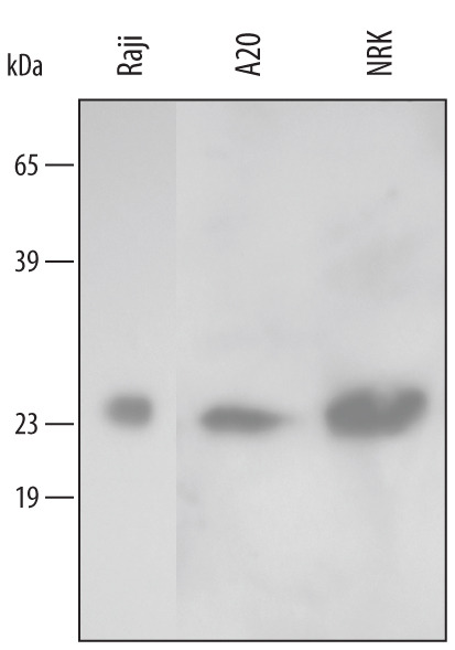
- Detection of Human/Mouse/Rat Peroxiredoxin 2 by Western Blot. Western blot shows lysates of Raji human Burkitt's lymphoma cell line, A20 mouse B cell lymphoma cell line, and NRK rat normal kidney cell line. PVDF membrane was probed with 0.2 µg/mL of Goat Anti-Human/Mouse/Rat Peroxiredoxin 2 Antigen Affinity-purified Polyclonal Antibody (Catalog # AF3489) followed by HRP-conjugated Anti-Goat IgG Secondary Antibody (Catalog # HAF109). A specific band was detected for Peroxiredoxin 2 at approximately 24 kDa (as indicated). This experiment was conducted using Immunoblot Buffer Group 2.
- Submitted by
- R&D Systems (provider)
- Main image

- Experimental details
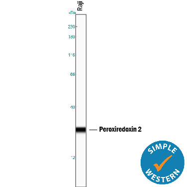
- Detection of Human Peroxiredoxin 2 by Simple WesternTM. Simple Western lane view shows lysates of Raji human Burkitt's lymphoma cell line, loaded at 0.2 mg/mL. A specific band was detected for Peroxiredoxin 2 at approximately 27 kDa (as indicated) using 2 µg/mL of Goat Anti-Human/Mouse/Rat Peroxiredoxin 2 Antigen Affinity-purified Polyclonal Antibody (Catalog # AF3489) followed by 1:50 dilution of HRP-conjugated Anti-Goat IgG Secondary Antibody (Catalog # HAF109). This experiment was conducted under reducing conditions and using the 12-230 kDa separation system.