Explore
 Validate
 Learn
Learn Western blot
 Immunocytochemistry
Antibody data
- Antibody Data
- Antigen structure
- References [2]
- Comments [0]
- Validations
- Immunocytochemistry [1]
- Immunohistochemistry [1]
- Other assay [3]
Submit
Validation data
Reference
Comment
Report error
- Product number
- PA5-30004 - Provider product page

- Provider
- Invitrogen Antibodies
- Product name
- CYP4A11 Polyclonal Antibody
- Antibody type
- Polyclonal
- Antigen
- Recombinant full-length protein
- Description
- Recommended positive controls: A549, HeLa, HepG2, HCT116. Predicted reactivity: Dog (88%), Bovine (83%). Store product as a concentrated solution. Centrifuge briefly prior to opening the vial.
- Reactivity
- Human
- Host
- Rabbit
- Isotype
- IgG
- Vial size
- 100 μL
- Concentration
- 1 mg/mL
- Storage
- Store at 4°C short term. For long term storage, store at -20°C, avoiding freeze/thaw cycles.
Submitted references Alteration of CYP4A11 expression in renal cell carcinoma: diagnostic and prognostic implications.
Cytochrome P450 4A11 expression in tumor cells: A favorable prognostic factor for hepatocellular carcinoma patients.
Kim S, Kim JM, Lee HJ, Lim JS, Seong IO, Kim KH
Journal of Cancer 2020;11(6):1478-1485
Journal of Cancer 2020;11(6):1478-1485
Cytochrome P450 4A11 expression in tumor cells: A favorable prognostic factor for hepatocellular carcinoma patients.
Eun HS, Cho SY, Lee BS, Kim S, Song IS, Chun K, Oh CH, Yeo MK, Kim SH, Kim KH
Journal of gastroenterology and hepatology 2019 Jan;34(1):224-233
Journal of gastroenterology and hepatology 2019 Jan;34(1):224-233
No comments: Submit comment
Supportive validation
- Submitted by
- Invitrogen Antibodies (provider)
- Main image

- Experimental details
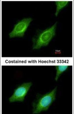
- Immunofluorescent analysis of CYP4A11 in methanol-fixed HeLa cells using a CYP4A11 polyclonal antibody (Product # PA5-30004) at a 1:200 dilution.
Supportive validation
- Submitted by
- Invitrogen Antibodies (provider)
- Main image

- Experimental details
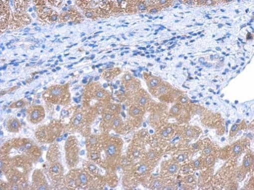
- Immunohistochemical analysis of paraffin-embedded human hepatoma, using CYP4A11 antibody (Product # PA5-30004) antibody at 1:500 dilution. Antigen Retrieval: EDTA based buffer, pH 8.0, 15 min.
Supportive validation
- Submitted by
- Invitrogen Antibodies (provider)
- Main image
- Experimental details
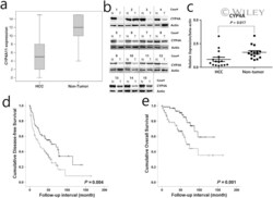
- 2 CYP4A11 protein expression and survival outcomes in hepatocellular carcinoma. (a) The 155 paired cases of hepatocellular carcinoma (HCC) and non-tumor tissue sections were assessed for CYP4A11 expression using the Wilcoxon signed-rank test ( P < 0.001). The line in the middle of the boxes is the median. The box length indicates the interquartile range. The ends of the whiskers represent maximum and minimum values. (b and c) Western blot analysis of CYP4A, which is homologous to human cytochrome P450 4A11 and 4A22, in 15 paired HCC and non-tumor tissue sections. The HCC tumor samples expressed significantly lower levels of CYP4A than the non-neoplastic samples ( P = 0.017; Wilcoxon signed-rank test). (b) Cell lysates were collected and subjected to western blot analysis for CYP4A. (c) Relative intensity of CYP4A protein expression in the HCC tumor and non-tumor tissue sections. Data are presented as mean +- SEM. (d and e) Kaplan-Meier survival curves for CYP4A11 expression in patients with HCC ( n = 155). The curves show significant associations between decreased CYP4A11 expression and shorter disease-free survival (log-rank = 8.390, P = 0.004) and overall survival (log-rank = 11.715, P < 0.001). (d and e) CYP4A11 expression: , low-expression; , high-expression; , low expression-censored; , high expression-censored.

- Submitted by
- Invitrogen Antibodies (provider)
- Main image
- Experimental details
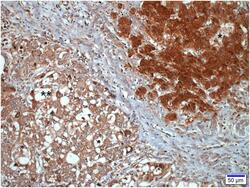
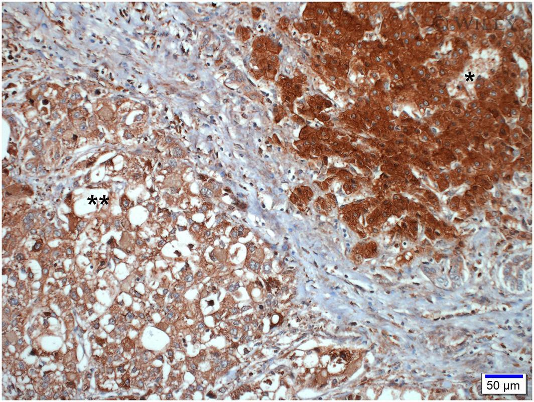
- 1 Representative photographs of CYP4A11 immunohistochemical staining in hepatocellular carcinoma. The tumor cells (**) show weak staining in contrast to the peritumoral non-neoplastic hepatocytes (*), which exhibit strong positive staining (scale bar = 50 mum).
- Submitted by
- Invitrogen Antibodies (provider)
- Main image

- Experimental details
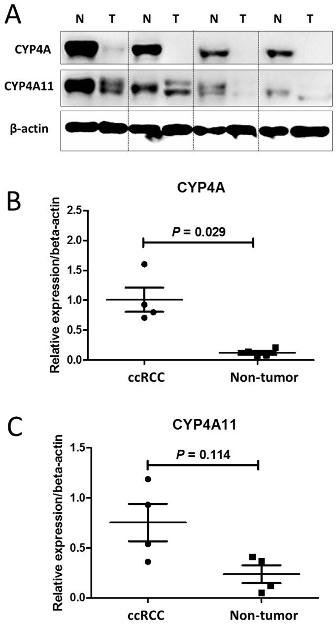
- Figure 2 Western blot analysis of CYP4A and CYP4A11 in 4 matched pairs of clear-cell renal cell carcinoma (ccRCC) tissue and nontumor tissue sections. The ccRCC tumor tissue samples expressed significantly lower levels of CYP4A and CYP4A11 than the nontumor tissue samples. (A) Cell lysates were collected and subjected to western blot analysis for CYP4A and CYP4A11. (B) Relative intensity of CYP4A protein expression in the ccRCC tumor and nontumor tissue sections (P = 0.029; Wilcoxon signed-rank test). (C) Relative intensity of CYP4A11 protein expression in the ccRCC tumor and nontumor tissue sections (P = 0.114; Wilcoxon signed-rank test).