Explore
 Validate
 Learn
Learn Western blot
 Immunocytochemistry
 Immunoprecipitation
Antibody data
- Antibody Data
- Antigen structure
- References [5]
- Comments [0]
- Validations
- Immunocytochemistry [1]
- Flow cytometry [1]
- Other assay [1]
Submit
Validation data
Reference
Comment
Report error
- Product number
- 32-1500 - Provider product page

- Provider
- Invitrogen Antibodies
- Product name
- Cyclin E Monoclonal Antibody (HE172)
- Antibody type
- Monoclonal
- Antigen
- Recombinant full-length protein
- Reactivity
- Human
- Host
- Mouse
- Isotype
- IgG
- Antibody clone number
- HE172
- Vial size
- 200 µg
- Concentration
- 0.5 mg/mL
- Storage
- -20°C
Submitted references Quantitative profiling of adaptation to cyclin E overproduction.
Caspases uncouple p27(Kip1) from cell cycle regulated degradation and abolish its ability to stimulate cell migration and invasion.
Evolutionary hypothesis of telomere length in primary breast cancer and brain tumour patients: a tracer for genomic-tumour heterogeneity and instability.
Molecular architecture of the DNA replication origin activation checkpoint.
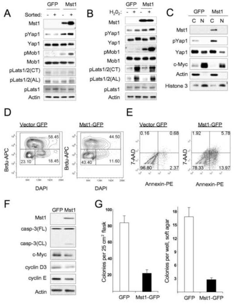
Mst1 and Mst2 maintain hepatocyte quiescence and suppress hepatocellular carcinoma development through inactivation of the Yap1 oncogene.
Limas JC, Littlejohn AN, House AM, Kedziora KM, Mouery BL, Ma B, Fleifel D, Walens A, Aleman MM, Dominguez D, Cook JG
Life science alliance 2022 May;5(5)
Life science alliance 2022 May;5(5)
Caspases uncouple p27(Kip1) from cell cycle regulated degradation and abolish its ability to stimulate cell migration and invasion.
Podmirseg SR, Jäkel H, Ranches GD, Kullmann MK, Sohm B, Villunger A, Lindner H, Hengst L
Oncogene 2016 Sep 1;35(35):4580-90
Oncogene 2016 Sep 1;35(35):4580-90
Evolutionary hypothesis of telomere length in primary breast cancer and brain tumour patients: a tracer for genomic-tumour heterogeneity and instability.
Mehdipour P, Kheirollahi M, Mehrazin M, Kamalian N, Atri M
Cell biology international 2011 Sep;35(9):915-25
Cell biology international 2011 Sep;35(9):915-25
Molecular architecture of the DNA replication origin activation checkpoint.
Tudzarova S, Trotter MW, Wollenschlaeger A, Mulvey C, Godovac-Zimmermann J, Williams GH, Stoeber K
The EMBO journal 2010 Oct 6;29(19):3381-94
The EMBO journal 2010 Oct 6;29(19):3381-94
Mst1 and Mst2 maintain hepatocyte quiescence and suppress hepatocellular carcinoma development through inactivation of the Yap1 oncogene.
Zhou D, Conrad C, Xia F, Park JS, Payer B, Yin Y, Lauwers GY, Thasler W, Lee JT, Avruch J, Bardeesy N
Cancer cell 2009 Nov 6;16(5):425-38
Cancer cell 2009 Nov 6;16(5):425-38
No comments: Submit comment
Supportive validation
- Submitted by
- Invitrogen Antibodies (provider)
- Main image
- Experimental details
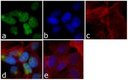
- Immunofluorescent analysis of Cyclin E was done on 70% confluent log phase HeLa cells. The cells were fixed with 4% paraformaldehyde for 15 minutes; permeabilized with 0.25% Triton™ X-100 for 10 minutes followed by blocking with 5% BSA for 1 hour at room temperature. The cells were incubated with Cyclin E Mouse Monoclonal Antibody (Product # 32-1500) at 2 µg - 4 µg in 1% BSA and incubated for 3 hours at room temperature and then labeled with Alexa Fluor® 488 Rabbit Anti-Mouse IgG Secondary Antibody (Product # A-11059) at a dilution of 1:400 for 30 minutes at room temperature (Panel a: green). Nuclei (Panel b: blue) were stained with SlowFade® Gold Antifade Mountant with DAPI (Product # S36938). F-actin (Panel c: red) was stained with Alexa Fluor® 594 Phalloidin (Product # A12381). Panel d is a merged image showing nuclear localization of Cyclin E. Panel e shows no primary antibody. The images were captured at 20X magnification.

Supportive validation
- Submitted by
- Invitrogen Antibodies (provider)
- Main image
- Experimental details
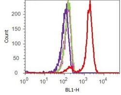
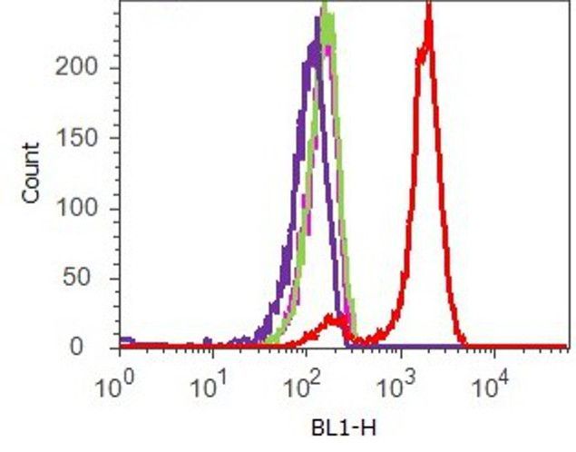
- Flow cytometry analysis of Cyclin E was done on synchronized HeLa cells. Cells were fixed with 70% ethanol for 10 minutes, permeabilized with 0.25% Triton™ X-100 for 20 minutes, and blocked with 5% BSA for 1 hour at room temperature. Cells were labeled with Cyclin E Mouse Monoclonal Antibody (321500, red histogram) or with mouse isotype control (pink histogram) at 2 µg - 4 µg/million cells in 2.5% BSA. After incubation at room temperature for 2 - 3 hours, the cells were labeled with Alexa Fluor® 488 Rabbit Anti-Mouse Secondary Antibody (A11059) at a dilution of 1:400 for 30 minutes at room temperature. The representative 10,000 cells were acquired and analyzed for each sample using an Attune® Acoustic Focusing Cytometer. The purple histogram represents unstained control cells and the green histogram represents no-primary-antibody control.
Supportive validation
- Submitted by
- Invitrogen Antibodies (provider)
- Main image
- Experimental details
- NULL