Explore
 Validate
 Learn
Learn32-1600
antibody from Invitrogen Antibodies
Targeting: CCNE1
CCNE
 Western blot
 Immunocytochemistry Immunoprecipitation Immunohistochemistry Flow cytometry Other assay
Antibody data
- Antibody Data
- Antigen structure
- References [13]
- Comments [0]
- Validations
- Immunocytochemistry [2]
- Immunohistochemistry [1]
- Flow cytometry [1]
- Other assay [2]
Submit
Validation data
Reference
Comment
Report error
- Product number
- 32-1600 - Provider product page

- Provider
- Invitrogen Antibodies
- Product name
- Cyclin E Monoclonal Antibody (HE12)
- Antibody type
- Monoclonal
- Antigen
- Recombinant full-length protein
- Reactivity
- Human
- Host
- Mouse
- Isotype
- IgG
- Antibody clone number
- HE12
- Vial size
- 200 μg
- Concentration
- 0.5 mg/mL
- Storage
- -20°C
Submitted references MicroRNA-18a targeting of the STK4/MST1 tumour suppressor is necessary for transformation in HPV positive cervical cancer.
A map of protein dynamics during cell-cycle progression and cell-cycle exit.
Immunohistochemical prediction of lapatinib efficacy in advanced HER2-positive breast cancer patients.
Suppression of apoptosis by PIF1 helicase in human tumor cells.
Neisseria gonorrhoeae infection induces altered amphiregulin processing and release.
A flow cytometry-based screen of nuclear envelope transmembrane proteins identifies NET4/Tmem53 as involved in stress-dependent cell cycle withdrawal.
Chemical genetics approach to restoring p27Kip1 reveals novel compounds with antiproliferative activity in prostate cancer cells.
E2F4 and ribonucleotide reductase mediate S-phase arrest in colon cancer cells treated with chlorophyllin.
Molecular classification and prognostication of adrenocortical tumors by transcriptome profiling.
Differential apoptosis by gallotannin in human colon cancer cells with distinct p53 status.
Neisseria gonorrhoeae infection causes a G1 arrest in human epithelial cells.
The F-box protein SKP2 mediates androgen control of p27 stability in LNCaP human prostate cancer cells.
The F-box protein SKP2 mediates androgen control of p27 stability in LNCaP human prostate cancer cells.
Morgan EL, Patterson MR, Ryder EL, Lee SY, Wasson CW, Harper KL, Li Y, Griffin S, Blair GE, Whitehouse A, Macdonald A
PLoS pathogens 2020 Jun;16(6):e1008624
PLoS pathogens 2020 Jun;16(6):e1008624
A map of protein dynamics during cell-cycle progression and cell-cycle exit.
Gookin S, Min M, Phadke H, Chung M, Moser J, Miller I, Carter D, Spencer SL
PLoS biology 2017 Sep;15(9):e2003268
PLoS biology 2017 Sep;15(9):e2003268
Immunohistochemical prediction of lapatinib efficacy in advanced HER2-positive breast cancer patients.
Duchnowska R, Wysocki PJ, Korski K, Czartoryska-Arłukowicz B, Niwińska A, Orlikowska M, Radecka B, Studziński M, Demlova R, Ziółkowska B, Merdalska M, Hajac Ł, Myśliwiec P, Zuziak D, Dębska-Szmich S, Lang I, Foszczyńska-Kłoda M, Karczmarek-Borowska B, Żawrocki A, Kowalczyk A, Biernat W, Jassem J, Central and East European Oncology Group (CEEOG)
Oncotarget 2016 Jan 5;7(1):550-64
Oncotarget 2016 Jan 5;7(1):550-64
Suppression of apoptosis by PIF1 helicase in human tumor cells.
Gagou ME, Ganesh A, Thompson R, Phear G, Sanders C, Meuth M
Cancer research 2011 Jul 15;71(14):4998-5008
Cancer research 2011 Jul 15;71(14):4998-5008
Neisseria gonorrhoeae infection induces altered amphiregulin processing and release.
Löfmark S, de Klerk N, Aro H
PloS one 2011 Jan 27;6(1):e16369
PloS one 2011 Jan 27;6(1):e16369
A flow cytometry-based screen of nuclear envelope transmembrane proteins identifies NET4/Tmem53 as involved in stress-dependent cell cycle withdrawal.
Korfali N, Srsen V, Waterfall M, Batrakou DG, Pekovic V, Hutchison CJ, Schirmer EC
PloS one 2011 Apr 14;6(4):e18762
PloS one 2011 Apr 14;6(4):e18762
Chemical genetics approach to restoring p27Kip1 reveals novel compounds with antiproliferative activity in prostate cancer cells.
Rico-Bautista E, Yang CC, Lu L, Roth GP, Wolf DA
BMC biology 2010 Dec 23;8:153
BMC biology 2010 Dec 23;8:153
E2F4 and ribonucleotide reductase mediate S-phase arrest in colon cancer cells treated with chlorophyllin.
Chimploy K, Díaz GD, Li Q, Carter O, Dashwood WM, Mathews CK, Williams DE, Bailey GS, Dashwood RH
International journal of cancer 2009 Nov 1;125(9):2086-94
International journal of cancer 2009 Nov 1;125(9):2086-94
Molecular classification and prognostication of adrenocortical tumors by transcriptome profiling.
Giordano TJ, Kuick R, Else T, Gauger PG, Vinco M, Bauersfeld J, Sanders D, Thomas DG, Doherty G, Hammer G
Clinical cancer research : an official journal of the American Association for Cancer Research 2009 Jan 15;15(2):668-76
Clinical cancer research : an official journal of the American Association for Cancer Research 2009 Jan 15;15(2):668-76
Differential apoptosis by gallotannin in human colon cancer cells with distinct p53 status.
Al-Ayyoubi S, Gali-Muhtasib H
Molecular carcinogenesis 2007 Mar;46(3):176-86
Molecular carcinogenesis 2007 Mar;46(3):176-86
Neisseria gonorrhoeae infection causes a G1 arrest in human epithelial cells.
Jones A, Jonsson AB, Aro H
FASEB journal : official publication of the Federation of American Societies for Experimental Biology 2007 Feb;21(2):345-55
FASEB journal : official publication of the Federation of American Societies for Experimental Biology 2007 Feb;21(2):345-55
The F-box protein SKP2 mediates androgen control of p27 stability in LNCaP human prostate cancer cells.
Lu L, Schulz H, Wolf DA
BMC cell biology 2002 Aug 20;3:22
BMC cell biology 2002 Aug 20;3:22
The F-box protein SKP2 mediates androgen control of p27 stability in LNCaP human prostate cancer cells.
Lu L, Schulz H, Wolf DA
BMC cell biology 2002 Aug 20;3:22
BMC cell biology 2002 Aug 20;3:22
No comments: Submit comment
Supportive validation
- Submitted by
- Invitrogen Antibodies (provider)
- Main image

- Experimental details
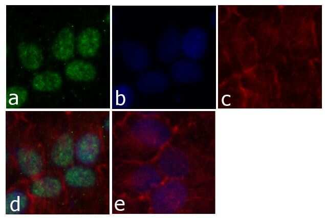
- Immunofluorescent analysis of Cyclin E was done on 70% confluent log phase HeLa cells. The cells were fixed with 4% paraformaldehyde for 15 minutes; permeabilized with 0.25% Triton™ X-100 for 10 minutes followed by blocking with 5% BSA for 1 hour at room temperature. The cells were incubated with Cyclin E Mouse Monoclonal Antibody (Product # 32-1600) at 2 µg - 4 µg in 1% BSA and incubated for 3 hours at room temperature and then labeled with Alexa Fluor® 488 Rabbit Anti-Mouse IgG Secondary Antibody (Product # A-11059) at a dilution of 1:400 for 30 minutes at room temperature (Panel a: green). Nuclei (Panel b: blue) were stained with SlowFade® Gold Antifade Mountant with DAPI (Product # S36938). F-actin (Panel c: red) was stained with Alexa Fluor® 594 Phalloidin (Product # A12381). Panel d is a merged image showing nuclear localization of Cyclin E. Panel e shows no primary antibody. The images were captured at 20X magnification.
- Submitted by
- Invitrogen Antibodies (provider)
- Main image

- Experimental details
- Immunofluorescent analysis of Cyclin E was done on 70% confluent log phase HeLa cells. The cells were fixed with 4% paraformaldehyde for 15 minutes; permeabilized with 0.25% Triton™ X-100 for 10 minutes followed by blocking with 5% BSA for 1 hour at room temperature. The cells were incubated with Cyclin E Mouse Monoclonal Antibody (Product # 32-1600) at 2 µg - 4 µg in 1% BSA and incubated for 3 hours at room temperature and then labeled with Alexa Fluor® 488 Rabbit Anti-Mouse IgG Secondary Antibody (Product # A-11059) at a dilution of 1:400 for 30 minutes at room temperature (Panel a: green). Nuclei (Panel b: blue) were stained with SlowFade® Gold Antifade Mountant with DAPI (Product # S36938). F-actin (Panel c: red) was stained with Alexa Fluor® 594 Phalloidin (Product # A12381). Panel d is a merged image showing nuclear localization of Cyclin E. Panel e shows no primary antibody. The images were captured at 20X magnification.

Supportive validation
- Submitted by
- Invitrogen Antibodies (provider)
- Main image
- Experimental details
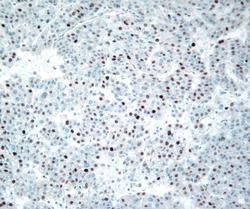
- Immunohistochemistry analysis of Cyclin E was done on hepatocellular carcinoma tissue. The tissue was probed with Cyclin E Mouse Monoclonal Antibody (Product # 32-1600).
Supportive validation
- Submitted by
- Invitrogen Antibodies (provider)
- Main image

- Experimental details
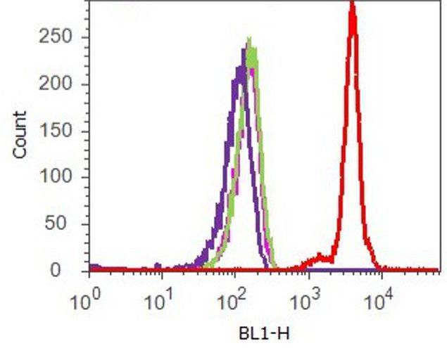
- Flow cytometry analysis of Cyclin E was done on synchronized HeLa cells. Cells were fixed with 70% ethanol for 10 minutes, permeabilized with 0.25% Triton™ X-100 for 20 minutes, and blocked with 5% BSA for 1 hour at room temperature. Cells were labeled with Cyclin E Mouse Monoclonal Antibody (321600, red histogram) or with mouse isotype control (pink histogram) at 2.5 µg/million cells in 2.5% BSA. After incubation at room temperature for 2 - 3 hours, the cells were labeled with Alexa Fluor® 488 Rabbit Anti-Mouse Secondary Antibody (A11059) at a dilution of 1:400 for 30 minutes at room temperature. The representative 10,000 cells were acquired and analyzed for each sample using an Attune® Acoustic Focusing Cytometer. The purple histogram represents unstained control cells and the green histogram represents no-primary-antibody control.
Supportive validation
- Submitted by
- Invitrogen Antibodies (provider)
- Main image
- Experimental details
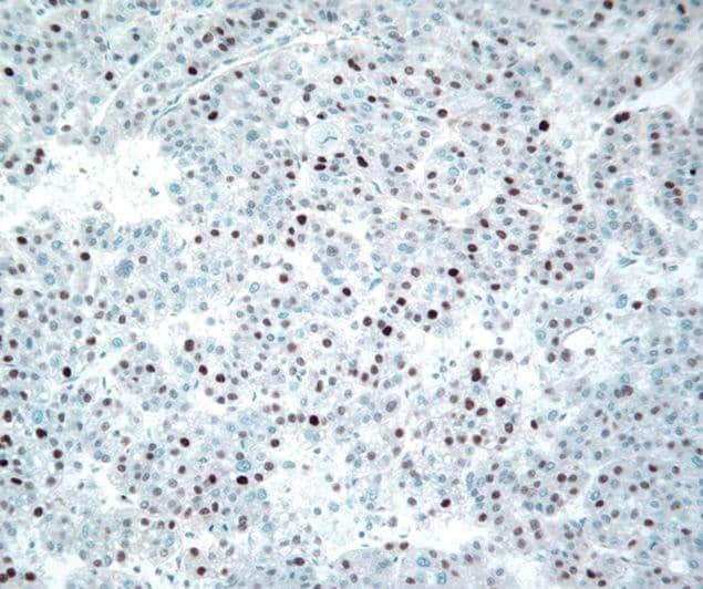
- NULL
- Submitted by
- Invitrogen Antibodies (provider)
- Main image
- Experimental details
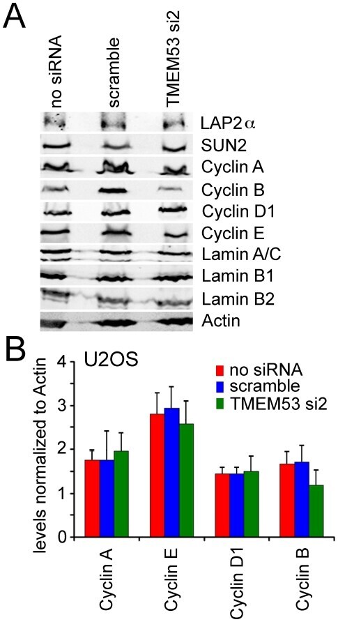
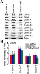
- Figure 9 NET4/TMEM53 knockdown did not affect levels of other nuclear envelope proteins linked to the cell cycle or cyclins. (A) Protein lysates were recovered from U2OS cells treated as in Figure 8 and reacted on Western blots with antibodies to the various proteins. This experiment was repeated 3 times and a representative Western blot is shown. (B) Cyclin levels were quantified from three separate experiments analyzed by LI-COR using fluorescent secondary antibodies and are plotted normalized to the actin control. Standard errors are shown.