Explore
 Validate
 Learn
LearnPA5-46983
antibody from Invitrogen Antibodies
Targeting: LILRB2
CD85d, ILT4, LIR-2, LIR2, MIR-10, MIR10
 Western blot Immunocytochemistry Immunohistochemistry
 Flow cytometry
 Blocking/Neutralizing Other assay
Antibody data
- Antibody Data
- Antigen structure
- References [1]
- Comments [0]
- Validations
- Western blot [1]
- Immunocytochemistry [2]
- Immunohistochemistry [2]
- Other assay [4]
Submit
Validation data
Reference
Comment
Report error
- Product number
- PA5-46983 - Provider product page

- Provider
- Invitrogen Antibodies
- Product name
- LILRB2 Polyclonal Antibody
- Antibody type
- Polyclonal
- Antigen
- Recombinant full-length protein
- Description
- In direct ELISAs and Western blots, approximately35% cross-reactivity with recombinant human (rh) ILT2 is observed and 5% cross-reactivity with rhILT5 is observed. Reconstitute at 0.2 mg/mL in sterile PBS. Endoxin level is
- Reactivity
- Human
- Host
- Goat
- Isotype
- IgG
- Vial size
- 100 μg
- Concentration
- 0.2 mg/mL
- Storage
- -20°C, Avoid Freeze/Thaw Cycles
Submitted references LILRB2-mediated TREM2 signaling inhibition suppresses microglia functions.
Zhao P, Xu Y, Jiang LL, Fan X, Ku Z, Li L, Liu X, Deng M, Arase H, Zhu JJ, Huang TY, Zhao Y, Zhang C, Xu H, Tong Q, Zhang N, An Z
Molecular neurodegeneration 2022 Jun 18;17(1):44
Molecular neurodegeneration 2022 Jun 18;17(1):44
No comments: Submit comment
Supportive validation
- Submitted by
- Invitrogen Antibodies (provider)
- Main image
- Experimental details
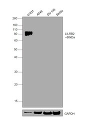
- Western Blot was performed using Anti-LILRB2 Polyclonal Antibody (Product # PA5-46983) and a 80 kDa band corresponding to Leukocyte immunoglobulin-like receptor subfamily B member 2 was observed in U-937 but not in A549, DU 145, BeWo. Whole cell extracts (30 µg lysate) of U-937 (Lane 1), A549 (Lane 2), DU 145 (Lane 3), BeWo (Lane 4) were electrophoresed using NuPAGE™ 10% Bis-Tris Protein Gel (Product # NP0302BOX). Resolved proteins were then transferred onto a nitrocellulose membrane (Product # IB23002) by iBlot® 2 Dry Blotting System (Product # IB21001). The blot was probed with the primary antibody (0.1 µg/mL) and detected by chemiluminescence with Rabbit anti-Goat IgG Heavy Chain Superclonal™ Recombinant Secondary Antibody, HRP (Product # A27014, 1/4000) using the iBright FL 1000 (Product # A32752). Chemiluminescent detection was performed using SuperSignal™ West Dura Extended Duration Substrate (Product # 34076).
Supportive validation
- Submitted by
- Invitrogen Antibodies (provider)
- Main image

- Experimental details
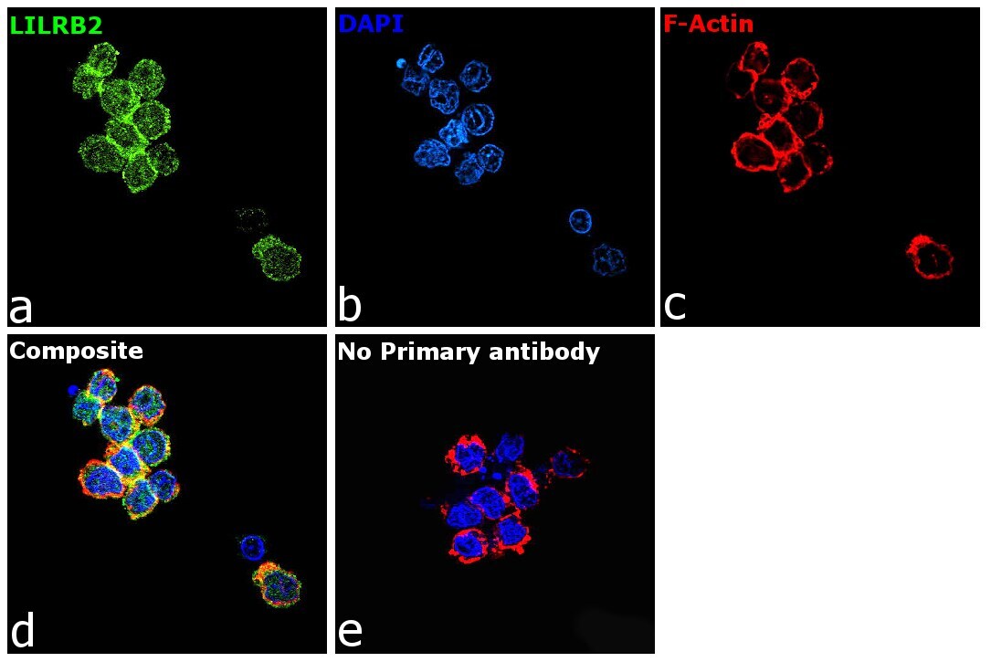
- Immunofluorescence analysis of Leukocyte immunoglobulin-like receptor subfamily B member 2 was performed using 70% confluent log phase U-937 cells. The cells were fixed with 4% paraformaldehyde for 10 minutes, permeabilized with 0.1% Triton™ X-100 for 15 minutes, and blocked with 2% BSA for 1 hour at room temperature. The cells were labeled with LILRB2 Polyclonal Antibody (Product # PA5-46983) at 5 µg/mL in 0.1% BSA, incubated at 4 degree celsius overnight and then labeled with Donkey anti-Goat IgG (H+L) Highly Cross-Adsorbed Secondary Antibody, Alexa Fluor Plus 488 (Product # A32814, 1/2000), for 45 minutes at room temperature (Panel a: Green). Nuclei (Panel b:Blue) were stained with ProLong™ Diamond Antifade Mountant with DAPI (Product # P36962). F-actin (Panel c: Red) was stained with Rhodamine Phalloidin (Product # R415, 1:300). Panel d represents the merged image showing membrane localization. Panel e represents control cells with no primary antibody to assess background. The images were captured at 60X magnification.

- Submitted by
- Invitrogen Antibodies (provider)
- Main image

- Experimental details
- Immunofluorescence analysis of Leukocyte immunoglobulin-like receptor subfamily B member 2 was performed using 70% confluent log phase U-937 cells. The cells were fixed with 4% paraformaldehyde for 10 minutes, permeabilized with 0.1% Triton™ X-100 for 15 minutes, and blocked with 2% BSA for 1 hour at room temperature. The cells were labeled with LILRB2 Polyclonal Antibody (Product # PA5-46983) at 5 µg/mL in 0.1% BSA, incubated at 4 degree celsius overnight and then labeled with Donkey anti-Goat IgG (H+L) Highly Cross-Adsorbed Secondary Antibody, Alexa Fluor Plus 488 (Product # A32814, 1/2000), for 45 minutes at room temperature (Panel a: Green). Nuclei (Panel b:Blue) were stained with ProLong™ Diamond Antifade Mountant with DAPI (Product # P36962). F-actin (Panel c: Red) was stained with Rhodamine Phalloidin (Product # R415, 1:300). Panel d represents the merged image showing membrane localization. Panel e represents control cells with no primary antibody to assess background. The images were captured at 60X magnification.
Supportive validation
- Submitted by
- Invitrogen Antibodies (provider)
- Main image
- Experimental details
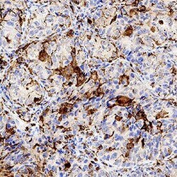
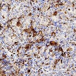
- Immunohistochemical analysis of LILRB2 in immersion fixed paraffin-embedded sections of human lung. Samples were incubated with LILRB2 polyclonal antibody (Product # PA5-46983) using a dilution of 1 µg/mL for 1 hour at room temperature followed by Anti-Goat IgG HRP Antibody. Tissue was stained using DAB (brown) and counterstained with hematoxylin (blue). Specific staining was localized to macrophages.

- Submitted by
- Invitrogen Antibodies (provider)
- Main image
- Experimental details
- Immunohistochemical analysis of LILRB2 in immersion fixed paraffin-embedded sections of human lung. Samples were incubated with LILRB2 polyclonal antibody (Product # PA5-46983) using a dilution of 1 µg/mL for 1 hour at room temperature followed by Anti-Goat IgG HRP Antibody. Tissue was stained using DAB (brown) and counterstained with hematoxylin (blue). Specific staining was localized to macrophages.

Supportive validation
- Submitted by
- Invitrogen Antibodies (provider)
- Main image
- Experimental details
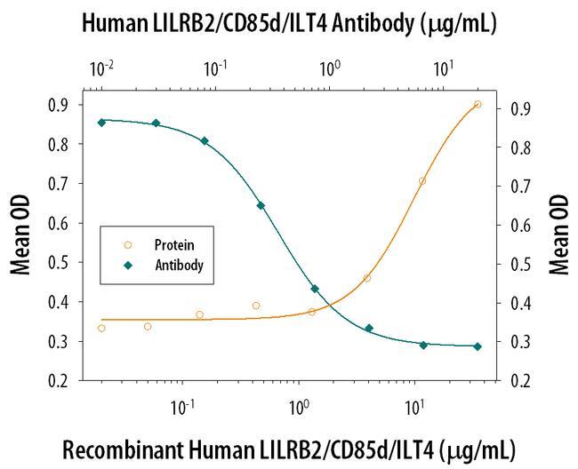
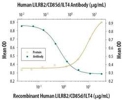
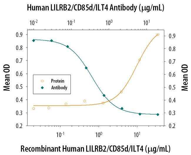
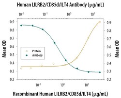
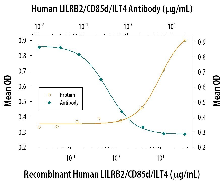
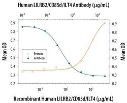
- Neutralization antibody testing demonstrates the specificity of an antibody through a correlation between antibody binding and the activity of the target. Neutralization of LILRB2 is shown by the decrease in mean OD (measure of adhesion of HSB2 cell line) with increasing concentrations of LILRB2 Polyclonal Antibody (PA5-46983).
- Submitted by
- Invitrogen Antibodies (provider)
- Main image
- Experimental details
- Neutralization of LILRB2 in HSB2 human peripheral blood acute lymphoblastic leukemia cell line. Samples were incubated in LILRB2 polyclonal antibody (Product # PA5-46983). Recombinant Human LILRB2/CD85d/ILT4 Fc Chimera, immobilized onto a microplate, supports the adhesion of the HSB2 human peripheral blood acute lymphoblastic leukemia cell line in a dose-dependent manner (orange line). Adhesion elicited by Recombinant Human LILRB2/CD85d/ILT4 Fc Chimera (35 µg/mL) is neutralized (green line) by increasing concentrations of Goat Anti-Human LILRB2/CD85d/ILT4 Antigen Affinity-purified Polyclonal Antibody. The ND50 is typically 0.3-1.0 µg/mL.
- Submitted by
- Invitrogen Antibodies (provider)
- Main image

- Experimental details
- Neutralization of LILRB2 in HSB2 human peripheral blood acute lymphoblastic leukemia cell line. Samples were incubated in LILRB2 polyclonal antibody (Product # PA5-46983). Recombinant Human LILRB2/CD85d/ILT4 Fc Chimera, immobilized onto a microplate, supports the adhesion of the HSB2 human peripheral blood acute lymphoblastic leukemia cell line in a dose-dependent manner (orange line). Adhesion elicited by Recombinant Human LILRB2/CD85d/ILT4 Fc Chimera (35 µg/mL) is neutralized (green line) by increasing concentrations of Goat Anti-Human LILRB2/CD85d/ILT4 Antigen Affinity-purified Polyclonal Antibody. The ND50 is typically 0.3-1.0 µg/mL.

- Submitted by
- Invitrogen Antibodies (provider)
- Main image
- Experimental details
- Neutralization antibody testing demonstrates the specificity of an antibody through a correlation between antibody binding and the activity of the target. Neutralization of LILRB2 is shown by the decrease in mean OD (measure of adhesion of HSB2 cell line) with increasing concentrations of LILRB2 Polyclonal Antibody (PA5-46983).