Explore
 Validate
 Learn
Learn12-5149-42
antibody from Invitrogen Antibodies
Targeting: LILRB2
CD85d, ILT4, LIR-2, LIR2, MIR-10, MIR10
 Flow cytometry
Antibody data
- Antibody Data
- Antigen structure
- References [6]
- Comments [0]
- Validations
- Flow cytometry [2]
- Other assay [5]
Submit
Validation data
Reference
Comment
Report error
- Product number
- 12-5149-42 - Provider product page

- Provider
- Invitrogen Antibodies
- Product name
- CD85d (ILT4) Monoclonal Antibody (42D1), PE, eBioscience™
- Antibody type
- Monoclonal
- Antigen
- Other
- Description
- Description: The monoclonal antibody 42D1 recognizes the CD85d protein also known as ILT4, LIR-2. It is a member of the ILT (immunoglobulin-like transcript)/ LIR (leukocyte Ig-like receptor), MIR (monocytes Ig-like receptor) family. CD85d is a 110 kDa single transmembrane receptor with 3 Ig-like domains and a long cytoplasmic domain containing ITIMs (immunoreceptor tyrosine-based inhibitory motifs). Expression is confined to monocytes, macrophages and dendritic cells. The ligand for CD85d is non-classical class I HLA-G, -B and G1. Upon ligand binding, tyrosine phosphatase SHP-1 interacts with the ITIM. Interaction of HLA-G with ILT4/CD85d is thought to contribute to tolerance during fetal implantation. Additionally, it is the levels of ILT3 and ILT4 on DCs that suggests a role in organ allograft rejection. Crosslinking of the receptor with the monoclonal antibody 42D1 inhibits calcium flux as well as enhancing HLA-G tetramer staining and restoring the stimulating activity of tolerogenic DC. Applications Reported: This 42D1 antibody has been reported for use in flow cytometric analysis. Applications Tested: This 42D1 antibody has been pre-titrated and tested by flow cytometric analysis of normal human peripheral blood cells. This can be used at 5 µL (1 µg) per test. A test is defined as the amount (µg) of antibody that will stain a cell sample in a final volume of 100 µL. Cell number should be determined empirically but can range from 10^5 to 10^8 cells/test. Excitation: 488-561 nm; Emission: 578 nm; Laser: Blue Laser, Green Laser, Yellow-Green Laser. Filtration: 0.2 µm post-manufacturing filtered.
- Reactivity
- Human
- Host
- Rat
- Conjugate
- Yellow dye
- Isotype
- IgG
- Antibody clone number
- 42D1
- Vial size
- 100 Tests
- Concentration
- 5 μL/Test
- Storage
- 4°C, store in dark, DO NOT FREEZE!
Submitted references Functionalization-dependent effects of cellulose nanofibrils on tolerogenic mechanisms of human dendritic cells.
Interferon beta and vitamin D synergize to induce immunoregulatory receptors on peripheral blood monocytes of multiple sclerosis patients.
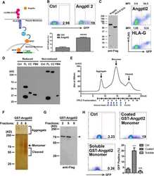
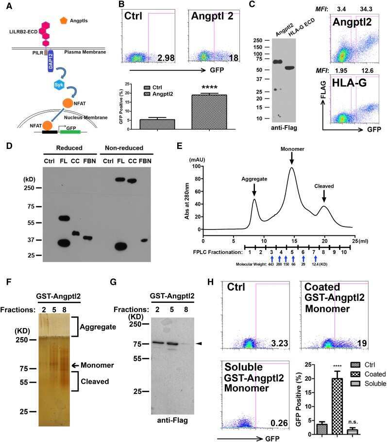
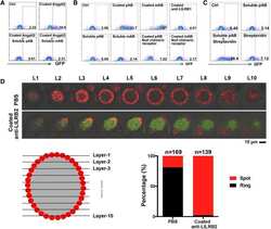
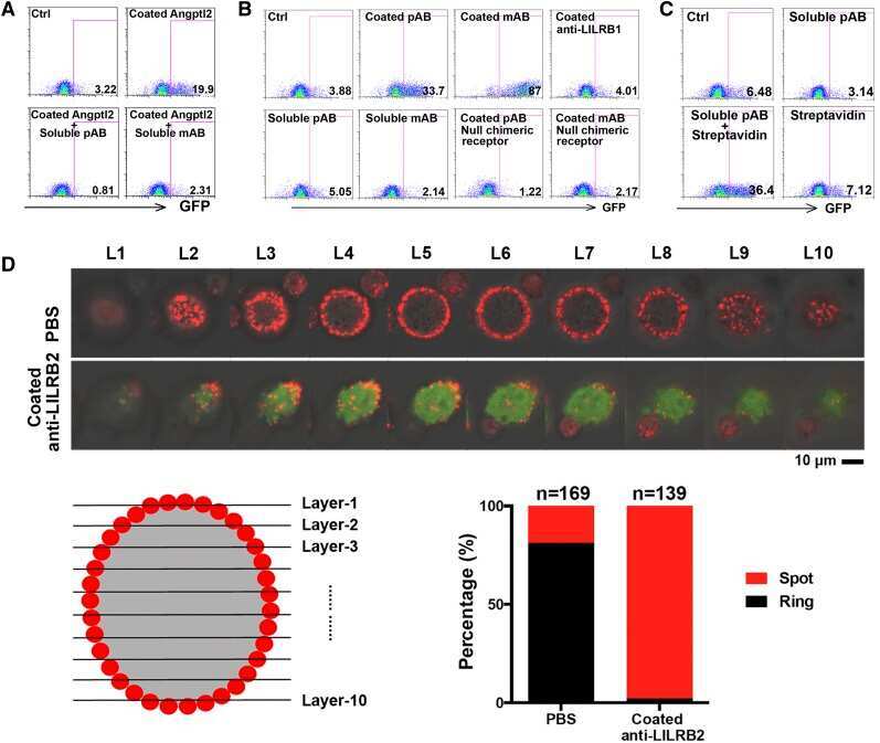
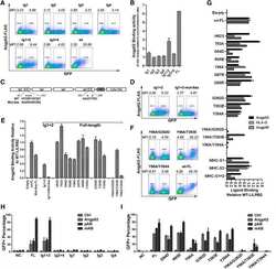
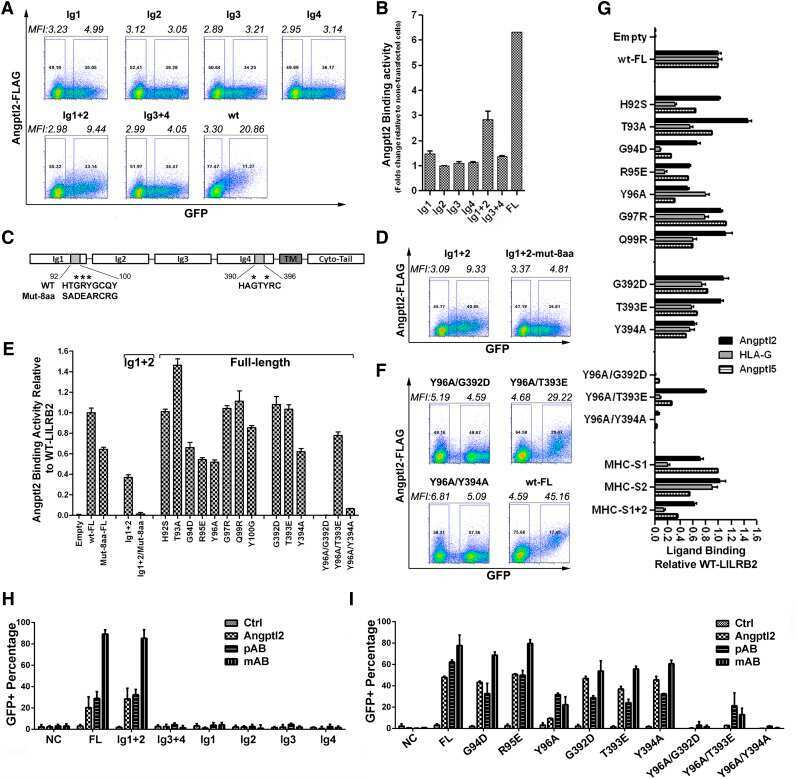
A motif in LILRB2 critical for Angptl2 binding and activation.
Mesenchymal stem cells from periapical lesions modulate differentiation and functional properties of monocyte-derived dendritic cells.
Inhibitory receptors bind ANGPTLs and support blood stem cells and leukaemia development.
High expression of ILT3 and ILT4 is a general feature of tolerogenic dendritic cells.
Tomić S, Ilić N, Kokol V, Gruden-Movsesijan A, Mihajlović D, Bekić M, Sofronić-Milosavljević L, Čolić M, Vučević D
International journal of nanomedicine 2018;13:6941-6960
International journal of nanomedicine 2018;13:6941-6960
Interferon beta and vitamin D synergize to induce immunoregulatory receptors on peripheral blood monocytes of multiple sclerosis patients.
Waschbisch A, Sanderson N, Krumbholz M, Vlad G, Theil D, Schwab S, Mäurer M, Derfuss T
PloS one 2014;9(12):e115488
PloS one 2014;9(12):e115488
A motif in LILRB2 critical for Angptl2 binding and activation.
Deng M, Lu Z, Zheng J, Wan X, Chen X, Hirayasu K, Sun H, Lam Y, Chen L, Wang Q, Song C, Huang N, Gao GF, Jiang Y, Arase H, Zhang CC
Blood 2014 Aug 7;124(6):924-35
Blood 2014 Aug 7;124(6):924-35
Mesenchymal stem cells from periapical lesions modulate differentiation and functional properties of monocyte-derived dendritic cells.
Dokić J, Tomić S, Marković M, Milosavljević P, Colić M
European journal of immunology 2013 Jul;43(7):1862-72
European journal of immunology 2013 Jul;43(7):1862-72
Inhibitory receptors bind ANGPTLs and support blood stem cells and leukaemia development.
Zheng J, Umikawa M, Cui C, Li J, Chen X, Zhang C, Huynh H, Kang X, Silvany R, Wan X, Ye J, Cantó AP, Chen SH, Wang HY, Ward ES, Zhang CC
Nature 2012 May 30;485(7400):656-60
Nature 2012 May 30;485(7400):656-60
High expression of ILT3 and ILT4 is a general feature of tolerogenic dendritic cells.
Manavalan JS, Rossi PC, Vlad G, Piazza F, Yarilina A, Cortesini R, Mancini D, Suciu-Foca N
Transplant immunology 2003 Jul-Sep;11(3-4):245-58
Transplant immunology 2003 Jul-Sep;11(3-4):245-58
No comments: Submit comment
Supportive validation
- Submitted by
- Invitrogen Antibodies (provider)
- Main image
- Experimental details
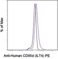
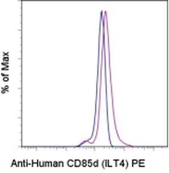
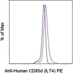
- Staining of normal human peripheral blood cells with Rat IgG2a K Isotype Control PE (Product # 12-4321-80) (blue histogram) or Anti-Human CD85d (ILT4) PE (purple histogram). Cells in the monocyte gate were used for analysis.
- Conjugate
- Yellow dye
- Submitted by
- Invitrogen Antibodies (provider)
- Main image
- Experimental details
- Staining of normal human peripheral blood cells with Rat IgG2a K Isotype Control PE (Product # 12-4321-80) (blue histogram) or Anti-Human CD85d (ILT4) PE (purple histogram). Cells in the monocyte gate were used for analysis.

Supportive validation
- Submitted by
- Invitrogen Antibodies (provider)
- Main image
- Experimental details
- NULL
- Conjugate
- Yellow dye
- Submitted by
- Invitrogen Antibodies (provider)
- Main image
- Experimental details
- NULL
- Conjugate
- Yellow dye
- Submitted by
- Invitrogen Antibodies (provider)
- Main image
- Experimental details
- NULL
- Conjugate
- Yellow dye
- Submitted by
- Invitrogen Antibodies (provider)
- Main image
- Experimental details
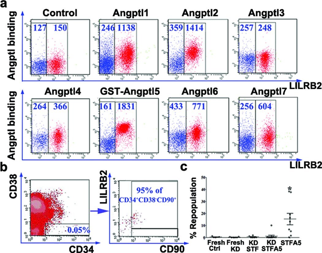
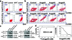
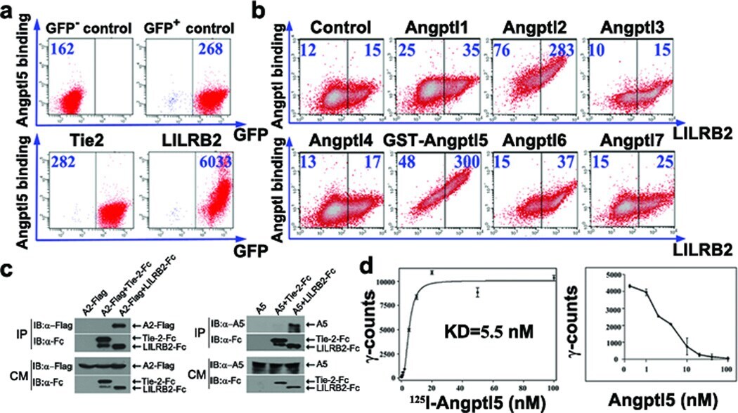
- Figure 1 Cell surface LILRB2 binds to Angptls a, Flow cytometry analysis of GST-Angptl5-FLAG binding to uninfected BAF3 cells or MSCV-GFP, MSCV-Tie2-GFP, or MSCV-LILRB2-GFP stably infected BAF3 cells. MFIs were indicated. b, Flow cytometry analysis of indicated FLAG-tagged Angptls binding to LILRB2 transfected 293T cells. c, Angptl2 and Angptl5 bound to the extracellular domain of LILRB2 but not Tie-2 in conditioned medium (CM) of co-transfected 293T cells (IB, immunoblotting; IP, immunoprecipitation). d-e, Concentration dependent specific ( d ) and competitive ( e ) 125 I-GST-Angptl5 binding to LILRB2 stably expressed BAF3 cells (n = 3). Error bars, s.e.m.
- Conjugate
- Yellow dye
- Submitted by
- Invitrogen Antibodies (provider)
- Main image
- Experimental details
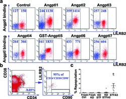
- Figure 2 LILRB2 mediates the effect of Angptl in supporting the repopulation of human cord blood HSCs a, Flow cytometry analysis of indicated FLAG-tagged Angptls binding to LILRB2 + human cord blood mononuclear cells (FACSAria). MFIs were indicated. b, Representative flow cytometry plots for the co-staining of CD34, CD38, CD90, and LILRB2 in human cord blood mononuclear cells (FACSCalibur). c, Human cord blood CD34 + cells infected with LILRB2 shRNA-encoding virus (KD) or control scramble shRNA virus were transplanted into sublethally irradiated NOD/SCID mice before or after culture for 10 d. SCF+TPO+Flt3L (STF) or STF+Angptl5 (STFA5) was used in the culture. Shown is the human donor repopulation after 2 months (n = 5-11). * p < 0.05. Error bars, s.e.m.
- Conjugate
- Yellow dye