Explore
 Validate
 Learn
Learn Western blot
 Immunocytochemistry
Antibody data
- Antibody Data
- Antigen structure
- References [2]
- Comments [0]
- Validations
- Immunocytochemistry [5]
- Other assay [4]
Submit
Validation data
Reference
Comment
Report error
- Product number
- PA5-20998 - Provider product page

- Provider
- Invitrogen Antibodies
- Product name
- ATG9B Polyclonal Antibody
- Antibody type
- Polyclonal
- Antigen
- Synthetic peptide
- Description
- A suggested positive control is Hela cell lysate. PA5-20998 can be used with blocking peptide PEP-1112.
- Reactivity
- Human, Mouse, Rat
- Host
- Rabbit
- Isotype
- IgG
- Vial size
- 100 μg
- Concentration
- 1 mg/mL
- Storage
- Maintain refrigerated at 2-8°C for up to 3 months. For long term storage store at -20°C
Submitted references Human papillomavirus 16E6/E7 activates autophagy via Atg9B and LAMP1 in cervical cancer cells.
Fisetin stimulates autophagic degradation of phosphorylated tau via the activation of TFEB and Nrf2 transcription factors.
Tingting C, Shizhou Y, Songfa Z, Junfen X, Weiguo L, Xiaodong C, Xing X
Cancer medicine 2019 Aug;8(9):4404-4416
Cancer medicine 2019 Aug;8(9):4404-4416
Fisetin stimulates autophagic degradation of phosphorylated tau via the activation of TFEB and Nrf2 transcription factors.
Kim S, Choi KJ, Cho SJ, Yun SM, Jeon JP, Koh YH, Song J, Johnson GV, Jo C
Scientific reports 2016 Apr 26;6:24933
Scientific reports 2016 Apr 26;6:24933
No comments: Submit comment
Supportive validation
- Submitted by
- Invitrogen Antibodies (provider)
- Main image

- Experimental details
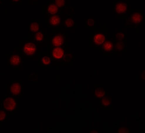
- Immunofluorescent analysis of HeLa cells using a ATG9B polyclonal antibody (Product # PA5-20998) at a 20 µg/mL dilution.
- Submitted by
- Invitrogen Antibodies (provider)
- Main image

- Experimental details
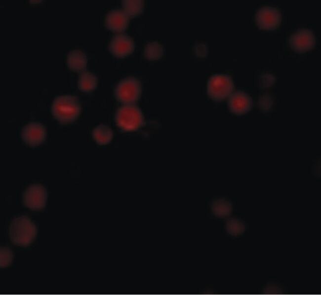
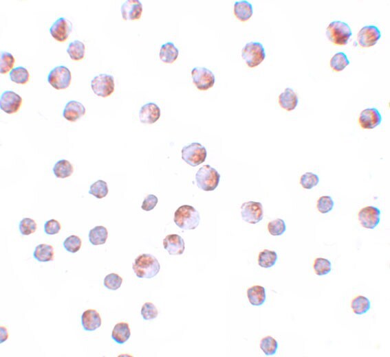
- Immunocytochemistry of ATG9B in HeLa cells with ATG9B Polyclonal Antibody (Product # PA5-20998) at 10 µg/mL.

- Submitted by
- Invitrogen Antibodies (provider)
- Main image

- Experimental details
- Immunofluorescence of ATG9B in Hela cells with ATG9B Polyclonal Antibody (Product # PA5-20998) at 20 µg/mL.
- Submitted by
- Invitrogen Antibodies (provider)
- Main image

- Experimental details
- Immunofluorescence of ATG9B in Hela cells with ATG9B Polyclonal Antibody (Product # PA5-20998) at 20 µg/mL.

- Submitted by
- Invitrogen Antibodies (provider)
- Main image
- Experimental details
- Immunocytochemistry of ATG9B in HeLa cells with ATG9B Polyclonal Antibody (Product # PA5-20998) at 10 µg/mL.
Supportive validation
- Submitted by
- Invitrogen Antibodies (provider)
- Main image

- Experimental details
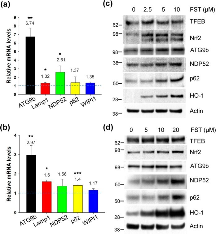
- Figure 6 Fisetin induces autophagy and lysosomal genes. ( a,b ) Mouse cortical cells (T4) and rat primary cortical neurons were treated with 5 and 10 muM fisetin for 24 h, respectively. Quantitative real time PCR (qRT-PCR) was performed using primer sets of genes of interest following the procedure described in Methods. Bar graphs represent the relative mRNA level of genes in T4 cells ( a ) and neurons ( b ) compared to those in cells not treated with fisetin. ( c,d ) T4 cells and neurons were treated with fisetin (FST) for 24 h and 36 h, respectively. The levels of TFEB, Nrf2, ATG9b, NDP52, p62/SQSTM1 and HO-1 in T4 cells ( c ) and neurons ( d ) were analyzed by immunoblotting using each corresponding antibody, respectively. Data shown are mean +- SE of three independent experiments and were analyzed using Student's t test. (* p < 0.05; ** p < 0.01; *** p < 0.001).
- Submitted by
- Invitrogen Antibodies (provider)
- Main image

- Experimental details
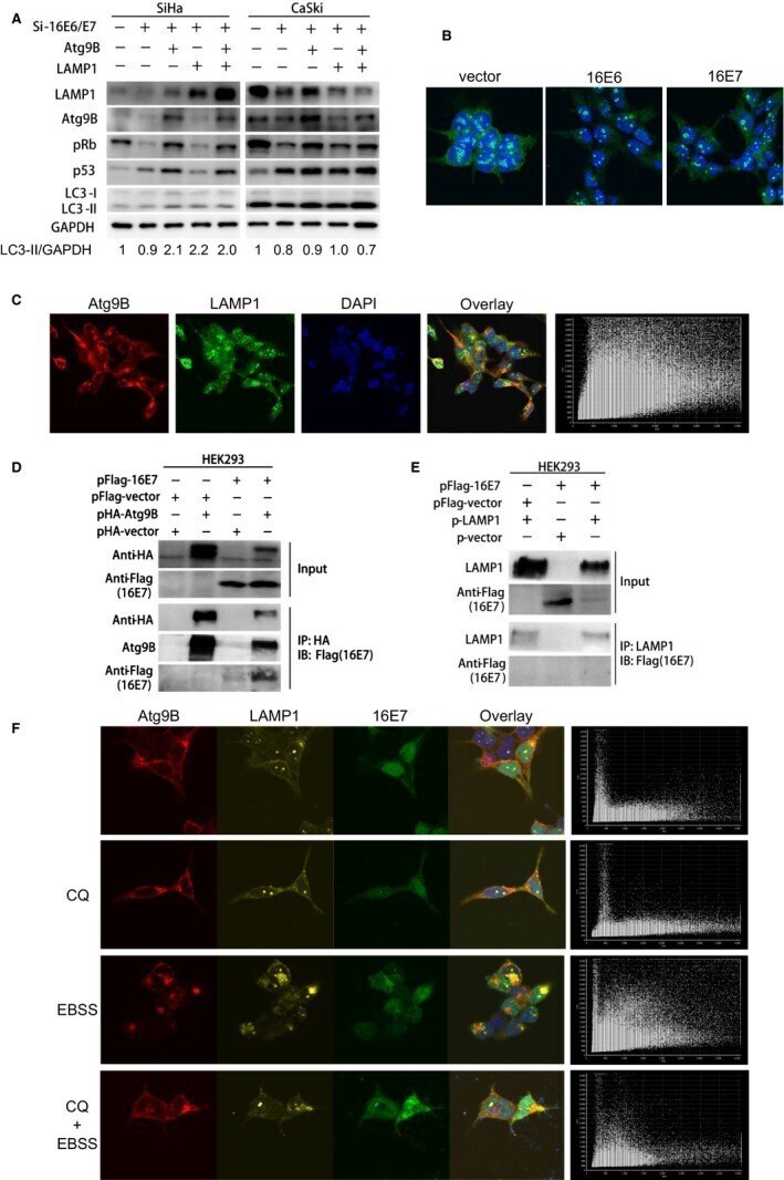
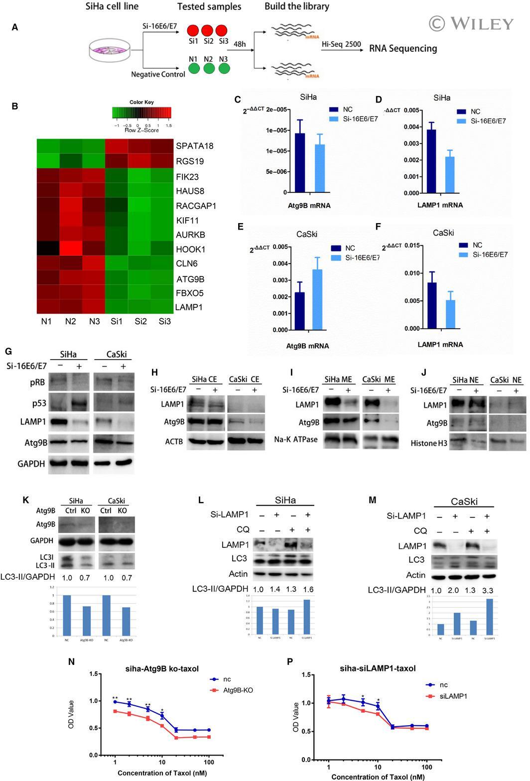
- Atg9B and LAMP1 were candidate genes involving in 16E6/E7 modulating autophagy. A, The process of RNA-sequencing. SiHa cells transfected with Si-16E6/E7 or negative RNAi (3 samples for each group) were prepared to build cDNA library. RNA-sequencing was performed in a Hiseq 2500 platform. B, Autophagy- and apoptosis-associated genes were selected to be candidate downstream regulators of 16E6/E7 and showed in a heatmap. C, The expression of Atg9B mRNA was downregulated in SiHa cells transfected with Si-16E6/E7. D, The expression of LAMP1 mRNA was downregulated in SiHa cells transfected with Si-16E7/E7. E, The expression of Atg9B mRNA was downregulated in CaSki cells transfected with Si-16E6/E7. F, The expression of LAMP1 mRNA was upregulated in CaSki cells transfected with Si-16E6/E7. G, The expression of LAMP1 and Atg9B protein was downregulated in SiHa and CaSki cells transfected with Si-16E6/E7. H, I, J, Atg9B, and LAMP1 were mainly detected in ME and decreased after 16E6/E7 knockdown in both SiHa and CaSki cells. K, LC3-II was decreased when Atg9B knockout by double nickase plasmid in SiHa and CaSki cells. L, LC3-II was accumulated when LAMP1 knockdown in SiHa cells with CQ exposure. M, LC3-II was accumulated when LAMP1 knockdown in CaSki cells, with or without CQ exposure. N, (P) Atg9B knockout or LAMP1 knockdown elevated the chemosensitivity of SiHa cells
- Submitted by
- Invitrogen Antibodies (provider)
- Main image

- Experimental details
- Figure 3 Atg9B and LAMP1 were candidate genes involving in 16E6/E7 modulating autophagy. A, The process of RNA-sequencing. SiHa cells transfected with Si-16E6/E7 or negative RNAi (3 samples for each group) were prepared to build cDNA library. RNA-sequencing was performed in a Hiseq 2500 platform. B, Autophagy- and apoptosis-associated genes were selected to be candidate downstream regulators of 16E6/E7 and showed in a heatmap. C, The expression of Atg9B mRNA was downregulated in SiHa cells transfected with Si-16E6/E7. D, The expression of LAMP1 mRNA was downregulated in SiHa cells transfected with Si-16E7/E7. E, The expression of Atg9B mRNA was downregulated in CaSki cells transfected with Si-16E6/E7. F, The expression of LAMP1 mRNA was upregulated in CaSki cells transfected with Si-16E6/E7. G, The expression of LAMP1 and Atg9B protein was downregulated in SiHa and CaSki cells transfected with Si-16E6/E7. H, I, J, Atg9B, and LAMP1 were mainly detected in ME and decreased after 16E6/E7 knockdown in both SiHa and CaSki cells. K, LC3-II was decreased when Atg9B knockout by double nickase plasmid in SiHa and CaSki cells. L, LC3-II was accumulated when LAMP1 knockdown in SiHa cells with CQ exposure. M, LC3-II was accumulated when LAMP1 knockdown in CaSki cells, with or without CQ exposure. N, (P) Atg9B knockout or LAMP1 knockdown elevated the chemosensitivity of SiHa cells

- Submitted by
- Invitrogen Antibodies (provider)
- Main image
- Experimental details
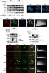
- Figure 4 Atg9B and LAMP1 are involved in 16E7 modulating autophagy in cervical cancer cells. A, LC3-II was accumulated when Atg9B were overexpressed and reduced when LAMP1 was overexpressed in 16E6/E7 knockdown cells, compared to the control group. B, LAMP1 was gathered as several clustered dots, while larger but less LAMP1 dots were formatted in HEK293 cells with overexpressed 16E6 or 16E7. C, Atg9B with red and LAMP1with green fluorescence were colocalized (Pearson's coefficient 0.726). D, 16E7 interacted with Atg9B. E, 16E7 do not interact with LAMP1. F, Atg9B was colocalized with LAMP1 in HEK293 cell with overexpressed 16E7 (Pearson's coefficient 0.457, 0.525, 0.717, and 0.456, respectively)